When we talk about Three-Dimensional Ultrasound Matrix Imaging, we’re diving into a technology that’s revolutionizing the way we see inside the human body. Imagine being able to look at your heart, not just as a flat image, but as a fully-formed, real-time 3D model. That’s what this groundbreaking technology offers. But before we get into the nitty-gritty of how it works, let’s set the scene with a little backstory.
The Evolution of Ultrasound
For decades, ultrasound has been a trusted tool in the medical field. You’ve probably seen those grainy, black-and-white images of babies in the womb or organs inside the body. These are 2D ultrasound images, which, while useful, provide a limited view. They’re like looking at a single page of a book instead of flipping through the whole thing. Now, think about what happens when you take those 2D images and stack them up, slice by slice. You get a three-dimensional view, something that gives you depth and a complete picture of what’s going on inside. Three-Dimensional Ultrasound Matrix Imaging takes this concept and runs with it, offering a much clearer, more detailed look at the body’s structures.
How Does Three-Dimensional Ultrasound Matrix Imaging Work?
So, how does Three-Dimensional Ultrasound Matrix Imaging actually work? Let’s break it down.
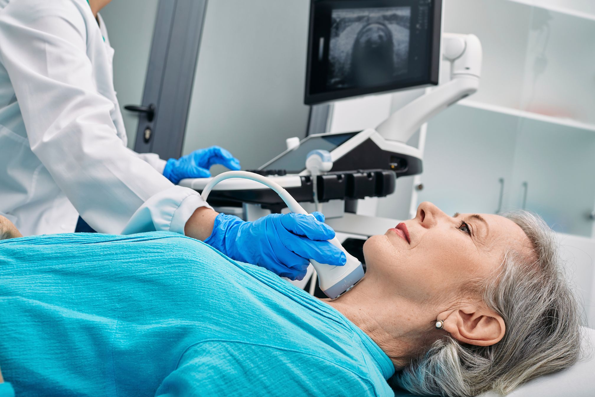
At the heart of this technology are matrix array transducers. Unlike traditional transducers, which send and receive ultrasound waves in a single plane, these matrix arrays have multiple rows and columns. Imagine a grid or matrix that can capture data from multiple angles at once. This allows for the creation of a volumetric dataset, which can then be rendered into a 3D image.
Here’s where it gets even more interesting. With Three-Dimensional Ultrasound Matrix Imaging, the data is captured in real-time. This means that instead of waiting for a technician to compile images, doctors can see the 3D images instantly. It’s like watching a live 3D movie of what’s happening inside your body.
Real-Life Applications: Where Does It Make a Difference?
Now, you might be wondering, “Where is Three-Dimensional Ultrasound Matrix Imaging actually used?” Great question! Let’s explore a few areas where this technology is making a real impact.
Cardiology: Seeing the Heart in 3D
One of the most exciting applications is in cardiology. The heart is a complex organ, constantly in motion, making it tricky to capture with traditional imaging. Three-Dimensional Ultrasound Matrix Imaging allows cardiologists to see the heart in action, in 3D, and from any angle. This means they can better assess heart function, detect abnormalities, and plan treatments more effectively.
For example, consider a patient with a suspected heart valve issue. Traditional 2D imaging might show the problem, but it doesn’t give the full picture. With Three-Dimensional Ultrasound Matrix Imaging, the cardiologist can view the valve in 3D, seeing exactly how it moves and interacts with surrounding tissues. This level of detail can make all the difference in diagnosis and treatment planning.
Obstetrics: A Clearer View of Fetal Development
In obstetrics, Three-Dimensional Ultrasound Matrix Imaging is giving parents and doctors a clearer view of fetal development. Traditional ultrasound images are often difficult for non-medical professionals to interpret, but 3D images are much easier to understand. Parents can see their baby’s face, limbs, and movements in incredible detail, making the experience even more special.
From a medical perspective, this technology allows for better monitoring of the baby’s development. Conditions like cleft palate or heart defects can be detected earlier and with more accuracy, allowing for timely interventions.
Oncology: Pinpointing Tumors with Precision
Cancer treatment is another area where Three-Dimensional Ultrasound Matrix Imaging shines. Tumors can be tricky to locate and even trickier to treat. But with this technology, doctors can get a more accurate view of where a tumor is located and how it’s interacting with surrounding tissues. This precision is crucial for planning surgeries or other treatments, minimizing damage to healthy tissue and improving patient outcomes.
The Benefits of Three-Dimensional Ultrasound Matrix Imaging
By now, it’s clear that Three-Dimensional Ultrasound Matrix Imaging is more than just a fancy new tool. It offers several key benefits that are transforming medical diagnostics.
Comprehensive Visualization
First and foremost, Three-Dimensional Ultrasound Matrix Imaging provides comprehensive visualization. Instead of seeing just one slice of the body, doctors can see the full picture. This is especially important in complex cases where a 2D image might miss something crucial.
Real-Time Data
Another major advantage is the real-time data it offers. In many medical situations, time is of the essence. Being able to view images instantly means that doctors can make faster, more informed decisions. Whether it’s during surgery or in an emergency room, this can be a lifesaver.
Non-Invasive and Safe
Just like traditional ultrasound, Three-Dimensional Ultrasound Matrix Imaging is non-invasive and safe. There’s no need for radiation or invasive procedures, making it a patient-friendly option for diagnostics.
Improved Diagnostic Accuracy
Finally, the accuracy of Three-Dimensional Ultrasound Matrix Imaging is unmatched. By providing a detailed, 3D view of the body’s structures, it helps doctors make more accurate diagnoses. This can lead to better treatment outcomes and, ultimately, better patient care.
Challenges and Future Directions
Of course, no technology is without its challenges, and Three-Dimensional Ultrasound Matrix Imaging is no exception. One of the main challenges is the cost. The equipment required for this technology is more expensive than traditional ultrasound machines, which can be a barrier for some medical facilities.
Another challenge is the need for specialized training. While the technology itself is user-friendly, interpreting 3D images requires a different skill set than 2D images. This means that healthcare professionals need additional training to use Three-Dimensional Ultrasound Matrix Imaging effectively.
That said, the future of Three-Dimensional Ultrasound Matrix Imaging looks bright. As the technology becomes more widespread and costs decrease, it’s likely to become a standard tool in medical diagnostics. Researchers are also exploring new applications for this technology, from improved cancer detection to better understanding of neurological conditions.
FAQ
What is a matrix ultrasound?
Matrix ultrasound is an advanced imaging technique that uses a grid-like array of transducers to capture data from multiple angles, producing more detailed, three-dimensional images compared to traditional ultrasound methods.
What is a three-dimensional diagnostic imaging?
Three-dimensional diagnostic imaging involves creating a 3D visual representation of internal body structures, allowing for a more comprehensive view than 2D imaging. It’s used for detailed examinations in fields like cardiology and obstetrics.
How does three-dimensional imaging work?
Three-dimensional imaging works by capturing multiple 2D images from different angles, which are then compiled into a 3D model. This allows for a more detailed and realistic view of the internal structures being examined.
Which technique results in a three-dimensional image ultrasound?
A three-dimensional image in ultrasound is typically achieved using matrix array transducers, which capture data from multiple planes simultaneously, allowing for the creation of a volumetric 3D image in real-time.
What are the 3 main types of ultrasound?
The three main types of ultrasound are 2D ultrasound, which provides flat images, 3D ultrasound, which gives a three-dimensional view, and Doppler ultrasound, which measures blood flow within vessels.
What is the difference between ultrasound and 3D ultrasound?
The difference between traditional ultrasound and 3D ultrasound lies in the image quality. While traditional ultrasound produces flat, 2D images, 3D ultrasound provides a more detailed and realistic three-dimensional view of the internal structures.
Why would a doctor order a 3D ultrasound?
A doctor might order a 3D ultrasound to get a clearer, more detailed view of the internal structures, especially for assessing fetal development, diagnosing heart conditions, or planning treatments for tumors.
What are the benefits of a 3D ultrasound?
The benefits of a 3D ultrasound include more detailed imaging, improved diagnostic accuracy, better visualization of complex structures, and the ability to view the images from multiple angles, which can enhance treatment planning.
Are 3D ultrasound images accurate?
Yes, 3D ultrasound images are highly accurate, providing detailed and realistic representations of internal structures. They offer better diagnostic clarity compared to 2D images, especially for complex cases
Conclusion
So, why should we care about Three-Dimensional Ultrasound Matrix Imaging? In short, because it’s changing the game in medical diagnostics. By offering a clearer, more detailed view of the body, this technology is helping doctors make better decisions, leading to better patient outcomes.
Whether it’s a cardiologist diagnosing a heart condition, an obstetrician monitoring a pregnancy, or an oncologist planning cancer treatment, Three-Dimensional Ultrasound Matrix Imaging is making a difference. And as the technology continues to evolve, its impact on the medical field is only going to grow.
In a world where precision and accuracy are paramount, Three-Dimensional Ultrasound Matrix Imaging stands out as a powerful tool for healthcare professionals. It’s not just about seeing inside the body; it’s about seeing it in a whole new way. And that’s something worth paying attention to.