When it comes to modern healthcare, abdominal ultrasound is a game-changer. You might not realize it, but this seemingly simple procedure holds the key to accurate diagnosis and comprehensive health assessment. From evaluating the liver to spotting kidney stones, abdominal ultrasound can do it all, providing detailed images of the body’s most vital organs without the need for surgery or exposure to harmful radiation.
Imagine you’re experiencing unexplained stomach pain. Instead of enduring invasive tests, an abdominal ultrasound offers a quick, painless way to get answers. But what exactly does this process involve, and why is it so important for your health? Let’s break it down.
What is an Abdominal Ultrasound?
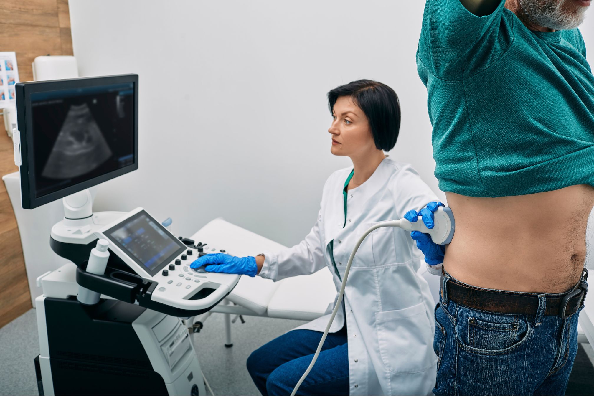
An abdominal ultrasound is a non-invasive imaging test that uses high-frequency sound waves to create pictures of the organs inside your abdomen. The process is simple: A technician applies a special gel to your skin and then uses a transducer (a small handheld device) to send sound waves into your body. These sound waves bounce off your organs and tissues, and the echoes are used to create detailed images on a monitor.
This test is often called precision imaging because it provides doctors with clear visuals of the abdomen’s key structures. Whether you’re concerned about gallstones, liver disease, or a potential pancreatic issue, an abdominal ultrasound offers a highly reliable assessment tool.
The Accuracy of Abdominal Ultrasound: Why Precision Matters
The term “precision imaging” isn’t just a fancy phrase—it reflects the superior accuracy of this technique. When your health is on the line, you want a diagnostic method that leaves no room for error. An abdominal ultrasound is capable of detecting a range of conditions with exceptional clarity.
For instance, if your doctor suspects you have gallstones, they can use an abdominal ultrasound to get a clear view of your gallbladder. This allows them to identify even the smallest stones, preventing unnecessary complications down the road. This level of accuracy is crucial for catching conditions early, offering you the best chance for successful treatment.
Real-Life Scenario: How Abdominal Ultrasound Helped Detect Liver Cirrhosis
Take the case of a 45-year-old man experiencing persistent fatigue and abdominal pain. Despite his symptoms, initial tests showed little cause for concern. It wasn’t until he had an abdominal ultrasound that doctors identified signs of liver cirrhosis—a serious condition that was progressing without obvious symptoms.
Thanks to the precision imaging of the abdominal ultrasound, the medical team was able to intervene before the condition became life-threatening. This example illustrates why an abdominal ultrasound is such a valuable tool for accurate diagnosis.
Comprehensive Health Assessment: What Can an Abdominal Ultrasound Detect?
You may be wondering: What exactly can an abdominal ultrasound detect? The answer is—quite a lot. This test provides a comprehensive health assessment by giving doctors insight into the functioning of several key organs. Here are some of the major organs evaluated during an abdominal ultrasound:
Liver
The liver is a vital organ responsible for filtering toxins from your bloodstream. An abdominal ultrasound can detect conditions like fatty liver disease, liver tumors, and cirrhosis. Given that liver problems often show no early symptoms, this kind of imaging can be life-saving.
Gallbladder
If you’ve ever had gallstones, you know how painful they can be. Fortunately, an abdominal ultrasound can quickly identify these stones, enabling your doctor to plan the best course of action.
Kidneys
Your kidneys play a crucial role in removing waste and excess fluid from your body. By using precision imaging, an abdominal ultrasound can detect kidney stones, cysts, and other abnormalities.
Pancreas
Although pancreatic issues can be hard to detect early, an abdominal ultrasound can help identify conditions like pancreatitis or pancreatic tumors, making it an essential part of any comprehensive health assessment.
Non-Invasive and Safe: Why Choose an Abdominal Ultrasound Over Other Methods?
One of the best things about an abdominal ultrasound is that it’s completely non-invasive. You don’t have to worry about needles, surgery, or long recovery times. It’s also incredibly safe, especially compared to procedures like CT scans or MRIs that expose you to radiation.
For pregnant women, children, or anyone who needs frequent imaging, the safety of an abdominal ultrasound makes it an excellent option. It’s also a much faster procedure, often taking less than 30 minutes from start to finish.
The Role of Abdominal Ultrasound in Monitoring Ongoing Conditions
An abdominal ultrasound isn’t just useful for one-time diagnoses. It also plays a crucial role in monitoring ongoing conditions. For example, if you have a known liver condition, your doctor might use regular ultrasounds to track its progression. This can help guide treatment plans and ensure you’re receiving the most effective care.
Similarly, patients with kidney disease or gallbladder issues can benefit from regular imaging. The ability to accurately diagnose changes in real time gives both patients and doctors peace of mind, knowing that they’re staying ahead of potential complications.
Common Uses: When Will Your Doctor Recommend an Abdominal Ultrasound?
Doctors commonly recommend abdominal ultrasounds for a variety of reasons. Some of the most common include:
- Unexplained Abdominal Pain: This is often the first step in figuring out the cause of pain in the abdomen, whether it’s gallstones, liver issues, or something else.
- Swelling or Bloating: If you’re experiencing unexplained bloating or swelling, an abdominal ultrasound can provide answers.
- Jaundice: Yellowing of the skin and eyes can indicate liver or bile duct issues, both of which are easily visible with an ultrasound.
- Kidney Stones: If you’re passing kidney stones, an abdominal ultrasound can determine the size and location of the stones.
Abdominal Ultrasound for Guided Procedures
Not only can an abdominal ultrasound be used for diagnosis, but it’s also extremely helpful for guiding certain medical procedures. For example, if your doctor needs to perform a biopsy (removing a small tissue sample for testing), they can use the ultrasound to guide the needle precisely, minimizing discomfort and risk.
Preparing for Your Abdominal Ultrasound
If your doctor has scheduled you for an abdominal ultrasound, don’t worry—it’s a simple process. Typically, you’ll be asked to fast for 8-12 hours before the procedure. This helps ensure the clearest images, as food and drink can interfere with the sound waves.
On the day of your ultrasound, you’ll lie comfortably on a table while the technician applies gel to your abdomen. The gel helps the transducer glide smoothly across your skin, ensuring clear images. You won’t feel any discomfort, and the whole process usually takes less than 30 minutes.
Conclusion
In today’s fast-paced healthcare environment, the need for precision imaging is greater than ever. Whether you’re experiencing unexplained symptoms or simply undergoing a routine health check, an abdominal ultrasound offers an unparalleled level of detail and accuracy.
From diagnosing liver conditions to detecting gallstones, the benefits of this non-invasive and safe procedure are hard to overstate. If your doctor suggests an abdominal ultrasound, you can trust that it’s a reliable and effective tool for ensuring your long-term health.
By providing a comprehensive health assessment, an abdominal ultrasound doesn’t just answer questions—it helps guide your entire healthcare journey