When it comes to non-invasive diagnostic tools, abdominal ultrasound is a go-to for many healthcare providers. The reason? It’s safe, effective, and provides real-time imaging that can be crucial in diagnosing a wide range of conditions. In this article, we’ll dive into the scanning techniques and interpretation methods that make abdominal ultrasound an indispensable part of modern medicine. Along the way, we’ll use real-life examples to make the content relatable and easy to understand.
Why Abdominal Ultrasound?
Imagine you’re a doctor faced with a patient who’s complaining of abdominal pain. There’s a myriad of potential causes—everything from gallstones to liver disease to kidney stones. An abdominal ultrasound offers a quick, painless way to start narrowing down the possibilities. It uses high-frequency sound waves to create images of the organs and structures within the abdomen, giving you a clear picture of what’s going on inside without having to resort to more invasive procedures.
Getting Started: The Importance of Preparation
Fasting: The Key to Clear Imaging
Before diving into the actual scanning techniques, let’s talk about preparation—because it can make all the difference in the clarity of the images. Most patients are asked to fast for 6-8 hours before the procedure. This might seem like a minor inconvenience, but it’s crucial. Fasting reduces the amount of gas in the intestines, which can otherwise obscure the view of the abdominal organs. It’s like trying to see through a foggy window—clearer conditions mean better results.
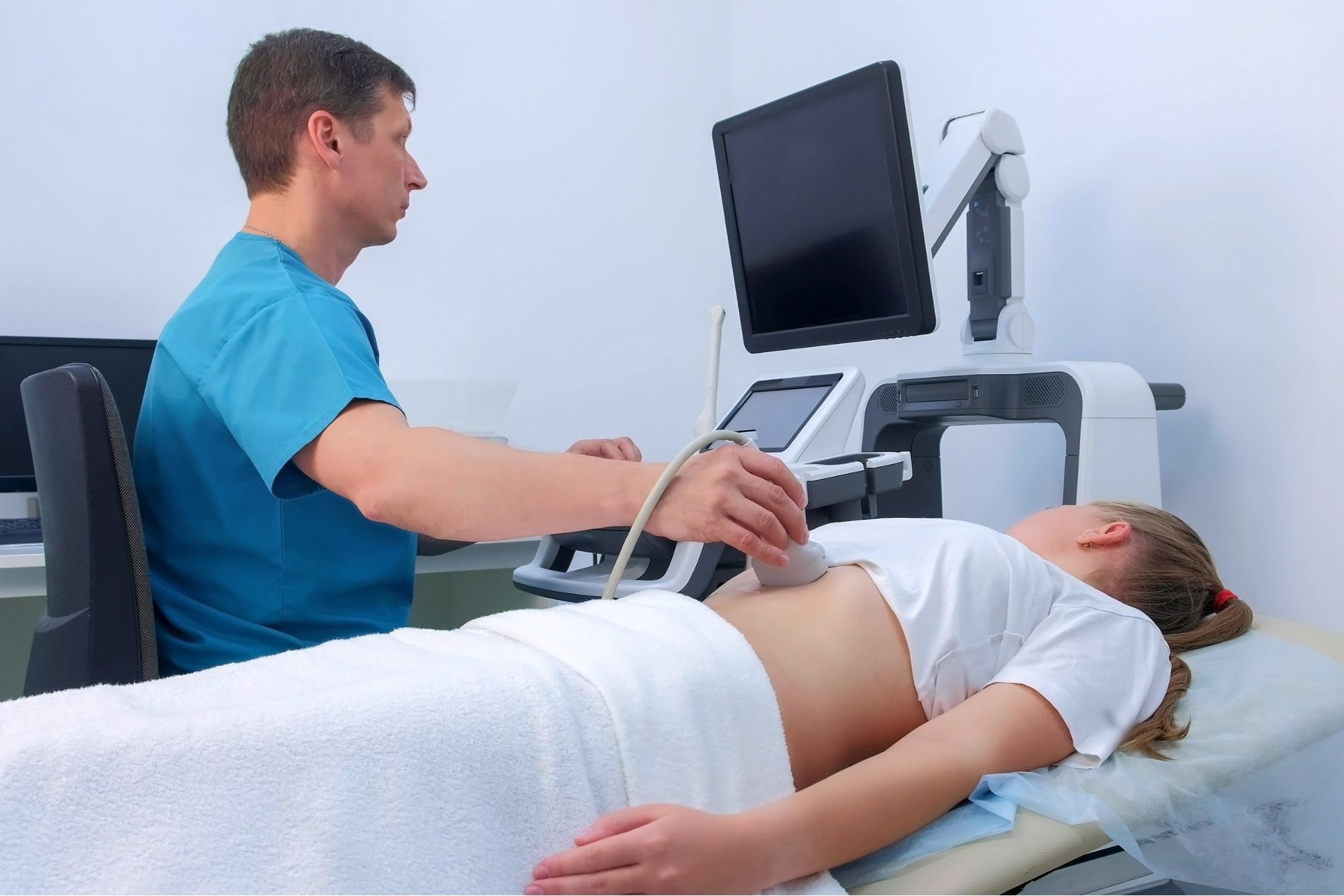
Patient Positioning: More Than Just Lying Down
Next up is patient positioning. You might think it’s as simple as lying down, but there’s actually some strategy involved. For a standard abdominal ultrasound, the patient is typically placed in a supine position—lying flat on their back. However, depending on which organ is being examined, the technician might ask the patient to lie on their left or right side. This can help bring certain organs, like the gallbladder or spleen, into a better view.
Scanning Techniques
Choosing the Right Transducer
One of the first decisions a sonographer has to make is selecting the appropriate transducer. The transducer is the device that sends and receives the sound waves. For abdominal ultrasound, a 3-5 MHz curvilinear transducer is usually the go-to choice. Lower frequencies can penetrate deeper tissues, making them ideal for imaging larger organs like the liver or kidneys. However, higher frequencies offer better resolution, which is crucial when you need to see finer details.
The Magic of Transducer Movements
Once the transducer is selected, the sonographer has to master a series of movements to capture the best images. These include sweeping, rocking, and sliding the transducer over the abdomen. Each movement helps to obtain different views of the organs. For example, rotating the transducer can provide multiple angles of the same structure, giving a more comprehensive picture. It’s a bit like taking a panorama shot with your camera—you want to capture as much detail as possible.
Interpreting the Images: What Are We Looking At?
The Liver: The Body’s Chemical Processing Plant
Let’s start with the liver—one of the most frequently examined organs in an abdominal ultrasound. A normal liver should appear homogenous, with a smooth surface and consistent echogenicity (brightness). But what if something doesn’t look quite right?
- Hepatomegaly, or an enlarged liver, could be a sign of liver disease, congestive heart failure, or even cancer.
- Focal lesions, which might show up as areas that are either brighter (hyperechoic) or darker (hypoechoic) than the surrounding tissue, could indicate anything from benign cysts to malignant tumors.
Imagine a patient with a history of alcohol abuse. During an abdominal ultrasound, the liver appears unusually bright—a classic sign of fatty liver disease. This finding can prompt further tests or interventions that might save the patient’s life.
The Gallbladder: Watch Out for Those Stones
Moving on to the gallbladder—this small, pear-shaped organ can cause big problems if things go wrong. A healthy gallbladder should appear as a dark (anechoic) structure with thin walls. However, one of the most common findings during an abdominal ultrasound is cholelithiasis, or gallstones. These show up as bright, echogenic spots with posterior acoustic shadowing—a fancy way of saying they cast a shadow on the ultrasound image.
Take, for example, a middle-aged woman who comes in with severe abdominal pain after eating a fatty meal. The ultrasound reveals multiple stones in her gallbladder, confirming the diagnosis of cholecystitis—inflammation of the gallbladder. This information is crucial in deciding whether she needs surgery.
The Pancreas: A Tricky Organ to Image
The pancreas can be a bit more challenging to image due to its location deep within the abdomen and behind other organs. However, with the right techniques, it’s possible to get a good look at this important organ. Normally, the pancreas should be slightly more echogenic than the liver and have a lobulated contour.
But what if the pancreas appears enlarged and has a heterogeneous texture? This could be a sign of pancreatitis—inflammation of the pancreas, which can be life-threatening if not treated promptly. Alternatively, a hypoechoic area within the pancreas might suggest a tumor, which could require further investigation or immediate intervention.
Other Key Structures: Spleen, Kidneys, and Aorta
The Spleen: The Body’s Blood Filter
The spleen is another organ that’s often examined during an abdominal ultrasound. A normal spleen should have a homogenous echotexture and be slightly more echogenic than the liver. However, an enlarged spleen (splenomegaly) might indicate underlying issues such as infections, blood disorders, or liver disease.
Consider a young adult with a history of frequent infections. During an ultrasound, the spleen is found to be significantly enlarged. This finding could prompt further tests to diagnose conditions like lymphoma or mononucleosis.
The Kidneys: The Body’s Waste Removal System
Kidneys are bean-shaped organs that play a crucial role in filtering waste from the blood. On an abdominal ultrasound, a normal kidney should have a hypoechoic cortex (the outer layer) and a central echogenic sinus (the inner part where urine collects).
However, if the renal pelvis and calyces (the structures where urine collects) appear dilated, this could indicate hydronephrosis—a condition where urine backs up into the kidney, often due to a blockage like a kidney stone. This is a common finding in patients who present with severe, sharp pain in the back or side.
The Aorta: The Body’s Main Artery
Last but not least, the abdominal aorta—the largest artery in the body. On ultrasound, it should appear as a tubular, anechoic structure with a pulsatile flow.
But what if you notice a localized dilation of the aorta? This could be an aortic aneurysm, a potentially life-threatening condition if the aneurysm ruptures. Detecting an aneurysm early through ultrasound can lead to life-saving interventions, such as surgery or endovascular repair.
Conclusion
By now, it’s clear that abdominal ultrasound is much more than just a tool—it’s a window into the body’s inner workings. From the liver to the kidneys, from the gallbladder to the pancreas, this non-invasive technique allows healthcare providers to diagnose and treat a wide range of conditions effectively. But what truly sets abdominal ultrasound apart is its ability to provide real-time imaging without the risks associated with radiation. It’s a testament to the power of sound waves and the skill of the sonographers who wield them. Whether you’re a patient seeking answers or a healthcare provider looking to make a diagnosis, abdominal ultrasound offers a safe, effective, and reliable solution.
So, the next time you hear someone mention abdominal ultrasound, you’ll know that it’s not just about capturing images—it’s about providing clarity in the quest for better health