Practical Applications of Ultrasound in Various Medical Fields
Ultrasound technology, a non-invasive imaging modality, has become an essential tool in modern medicine. With its ability to provide real-time images of soft tissues, organs, and blood flow, ultrasound plays a critical role in diagnosing and managing a wide range of conditions across different medical fields. Its versatility, safety, and relatively low cost make it indispensable for clinicians. This article explores the practical applications of ultrasound in various medical disciplines and how it enhances patient care.
1. Obstetrics and Gynecology
One of the most recognized uses of ultrasound is in obstetrics and gynecology. Ultrasound allows for the detailed visualization of the fetus during pregnancy. It is used to monitor fetal growth, detect congenital anomalies, confirm gestational age, and assess the placenta and amniotic fluid. The development of 3D and 4D ultrasound has further enhanced fetal imaging by allowing detailed views of fetal anatomy and real-time visualization of fetal movements.
In gynecology, ultrasound is crucial for diagnosing conditions like ovarian cysts, uterine fibroids, and endometriosis. It aids in the assessment of the pelvic organs, including the uterus, ovaries, and fallopian tubes, helping in the diagnosis and treatment planning for various reproductive health issues.
2. Cardiology
In cardiology, ultrasound, specifically echocardiography, is a vital tool for assessing heart function. It provides real-time images of the heart’s structure and motion, allowing cardiologists to evaluate the heart’s chambers, valves, and blood flow. Echocardiography is used to diagnose conditions such as heart valve disease, congenital heart defects, cardiomyopathy, and heart failure.
Doppler ultrasound, which measures the direction and speed of blood flow, is used to assess cardiac blood flow and detect issues like valve regurgitation or stenosis. Transesophageal echocardiography (TEE) offers a more detailed view of the heart by inserting a probe into the esophagus, providing high-quality images of the heart’s structures, especially in patients with difficult imaging windows due to obesity or lung disease.
3. Musculoskeletal Medicine
Ultrasound has become increasingly popular in musculoskeletal (MSK) medicine. It allows for the dynamic assessment of joints, tendons, ligaments, and muscles, providing crucial information for diagnosing injuries such as rotator cuff tears, tendonitis, and ligament sprains. It is also used for guiding therapeutic injections, such as corticosteroids, into affected areas with precision, ensuring that treatments are delivered accurately.
In addition, ultrasound is used to evaluate soft tissue masses, detect fluid collections like abscesses, and guide biopsies or aspirations. Its ability to provide real-time imaging during movement makes it particularly useful in assessing conditions like carpal tunnel syndrome or sports-related injuries.
4. Abdominal Imaging
In the realm of abdominal imaging, ultrasound is used to examine the liver, gallbladder, pancreas, kidneys, spleen, and other abdominal organs. It is especially useful for detecting gallstones, liver cirrhosis, abdominal aortic aneurysms, and kidney stones. Doppler ultrasound is also employed to assess blood flow in abdominal vessels, such as the hepatic and portal veins, helping diagnose conditions like portal hypertension or thrombosis.
Because of its safety and effectiveness, ultrasound is often the first-line imaging choice for evaluating patients with abdominal pain. It is also commonly used in emergency settings to quickly assess trauma patients for internal bleeding or organ damage.
5. Vascular Ultrasound
Vascular ultrasound is a crucial tool in evaluating blood flow in the body’s arteries and veins. It is used to diagnose conditions such as deep vein thrombosis (DVT), peripheral artery disease (PAD), and carotid artery stenosis. By using Doppler ultrasound to assess blood flow, clinicians can detect blockages, narrowing, or clots in the blood vessels.
Vascular ultrasound is particularly important in detecting DVT, a potentially life-threatening condition that can lead to pulmonary embolism if not treated promptly. It is also used to monitor the success of vascular surgeries, such as bypass grafts or stent placements, by evaluating blood flow and vessel patency postoperatively.
6. Emergency Medicine
In emergency medicine, ultrasound is a vital tool due to its speed, portability, and real-time imaging capabilities. Point-of-care ultrasound (POCUS) is used by emergency physicians to quickly assess patients in critical situations. It helps diagnose life-threatening conditions such as internal bleeding, pneumothorax, pericardial effusion, and abdominal aortic aneurysms.
POCUS is also used to guide emergency procedures such as central line placement, thoracentesis, and paracentesis. Its ability to provide immediate, bedside information significantly improves the speed and accuracy of diagnoses in emergency settings, often determining the course of treatment.
7. Breast Imaging
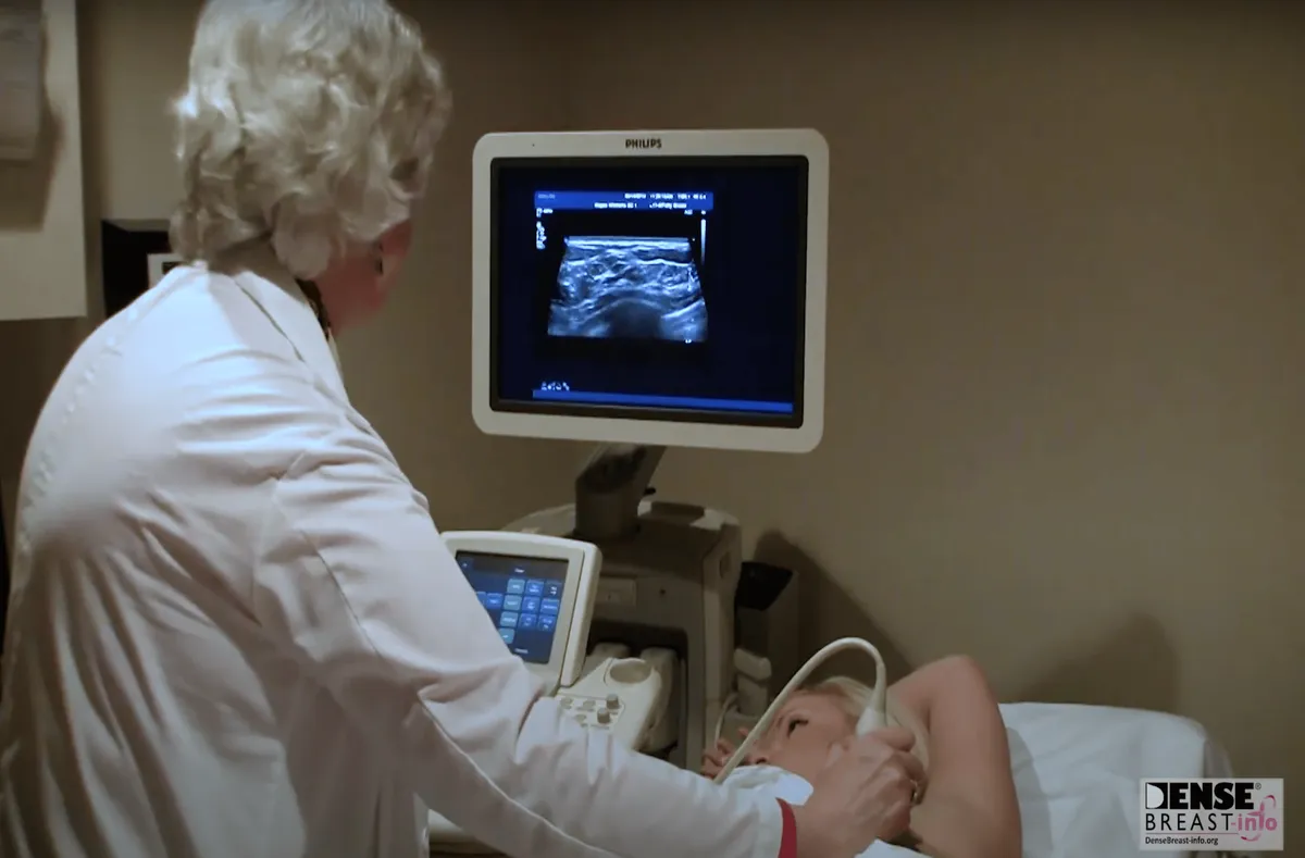
Ultrasound is commonly used in breast imaging to evaluate abnormalities found during mammography or physical examination. It is particularly useful for differentiating between solid masses and cysts and for guiding needle biopsies. Ultrasound is often the preferred imaging modality for younger women with dense breast tissue, where mammography may be less effective.
Breast ultrasound plays a crucial role in early detection of breast cancer, aiding in the characterization of suspicious lumps and ensuring timely biopsy or intervention. It is also used in follow-up imaging to monitor patients after surgery or during cancer treatment.
8. Urology
In urology, ultrasound is used to evaluate the kidneys, bladder, and prostate. It is commonly employed to detect kidney stones, bladder tumors, and prostate enlargement. Transrectal ultrasound (TRUS) is a specific type of ultrasound used to image the prostate gland, often utilized in the diagnosis and management of prostate cancer.
Additionally, urologists use ultrasound to guide minimally invasive procedures, such as needle biopsies of the prostate, ensuring accurate tissue sampling with minimal discomfort for the patient.
9. Gastroenterology
Ultrasound also has applications in gastroenterology, particularly in the diagnosis of liver diseases. It is used to assess liver size, detect liver tumors, and monitor conditions such as fatty liver disease or cirrhosis. Endoscopic ultrasound (EUS) is a specialized technique combining endoscopy and ultrasound to provide detailed images of the digestive tract and surrounding structures.
EUS is particularly useful for evaluating pancreatic and biliary disorders, staging gastrointestinal cancers, and guiding fine-needle aspirations or biopsies for diagnostic purposes.
10. Pediatrics
Pediatric ultrasound is used for a wide range of diagnostic purposes, from assessing congenital abnormalities in newborns to evaluating abdominal pain in children. It is a preferred imaging technique for children because it does not involve radiation and is non-invasive.
Neonatal brain ultrasound is commonly performed on premature infants to assess brain development and detect conditions like intraventricular hemorrhage. In addition, ultrasound is used to evaluate hip dysplasia in infants and guide treatment decisions.
FAQ
What is ultrasound commonly used for in obstetrics?
It is used to monitor fetal growth, detect congenital anomalies, and assess the placenta and amniotic fluid.
How does ultrasound help in cardiology?
It helps assess heart function, diagnose valve diseases, and evaluate cardiac blood flow using echocardiography.
What is the role of ultrasound in musculoskeletal medicine?
It helps diagnose injuries such as rotator cuff tears and guide therapeutic injections.
Which abdominal conditions can ultrasound detect?
It detects conditions like gallstones, liver cirrhosis, and abdominal aortic aneurysms.
How is vascular ultrasound beneficial?
It diagnoses conditions like deep vein thrombosis and peripheral artery disease by assessing blood flow in vessels.
Why is ultrasound important in emergency medicine?
It provides rapid, real-time imaging to diagnose conditions like internal bleeding or pneumothorax.
What role does ultrasound play in breast imaging?
It helps differentiate between solid masses and cysts and guides needle biopsies.
How is ultrasound used in urology?
It detects kidney stones, bladder tumors, and prostate enlargement and guides biopsies.
What is endoscopic ultrasound used for?
It evaluates pancreatic and biliary disorders and helps stage gastrointestinal cancers.
Why is ultrasound preferred for pediatric imaging?
It is non-invasive, safe, and does not involve radiation.
Conclusion
Ultrasound technology has become a cornerstone of medical imaging, offering valuable insights across a wide range of specialties. Its versatility, safety, and diagnostic accuracy make it an invaluable tool in both routine and emergency care. From obstetrics and cardiology to musculoskeletal medicine and emergency care, the practical applications of ultrasound continue to grow, improving patient outcomes and enabling more precise and timely interventions.


