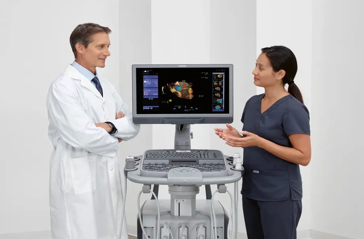
Differentiating Between Normal and Abnormal Findings
Ultrasound imaging is a powerful diagnostic tool that provides real-time, non-invasive visualization of the internal structures of the body. It is widely used in various medical fields, including obstetrics, cardiology, musculoskeletal imaging, and oncology, to assess a wide range of conditions. However, the key to using ultrasound effectively lies in the ability to differentiate between normal and abnormal findings. Accurate interpretation of ultrasound images is essential for ensuring proper diagnosis, treatment, and patient care.
In this article, we will explore how to distinguish between normal and abnormal ultrasound findings, focusing on the characteristics of healthy anatomy versus pathology, and the importance of experience and training in making accurate assessments.
Understanding Normal Ultrasound Findings
Before identifying abnormal ultrasound findings, it is essential to understand what constitutes a normal ultrasound image. Normal ultrasound results typically show anatomical structures that are healthy and free from disease. These structures have expected size, shape, and echogenicity (the ability to reflect ultrasound waves). A good understanding of what constitutes “normal” anatomy forms the foundation for identifying abnormalities.
Key Characteristics of Normal Ultrasound Findings
- Clear and Well-Defined Structures
In a normal ultrasound, anatomical structures should appear well-defined, with clear boundaries. Organs like the liver, kidneys, and heart should be visible with sharp edges and regular contours. - Appropriate Size and Shape
The size of an organ or structure should be within the expected range. For example, a normal-sized liver will appear smooth and even without any enlargement, while a heart should have a typical size and shape based on the patient’s age and sex. - Symmetry
Many paired organs, such as kidneys or ovaries, should be symmetrical. Asymmetry can be a sign of a pathological condition, such as a mass or abnormal growth. - Homogeneous Echogenicity
Healthy tissues show uniform echogenicity, meaning they reflect ultrasound waves evenly. For instance, the liver and kidneys should have a consistent echo pattern, and areas of increased or decreased echogenicity may suggest underlying pathology. - Absence of Abnormalities
A normal ultrasound image shows no evidence of masses, cysts, fluid collections, or abnormal blood flow. Any deviation from the typical appearance could indicate pathology. - Normal Doppler Flow
When using Doppler ultrasound to assess blood flow, normal results show smooth, regular flow patterns in blood vessels, without any blockages or irregularities.
Identifying Abnormal Ultrasound Findings
When ultrasound images reveal abnormalities, it’s important to interpret these findings carefully. Abnormal ultrasound findings often indicate disease or injury and can vary widely in terms of severity. The challenge lies in identifying these abnormalities early, which is essential for timely intervention.
Key Characteristics of Abnormal Ultrasound Findings
- Presence of Masses or Lesions
One of the most concerning ultrasound findings is the appearance of abnormal masses or lesions. These could represent benign growths, such as cysts or tumors, or malignant conditions like cancer. Masses may appear as solid or cystic and may require further investigation through biopsy or additional imaging. - Fluid Collections or Cysts
Fluid-filled structures, such as cysts or abscesses, are often visible on ultrasound. While simple cysts may be benign, complex cysts or fluid collections may indicate infection, malignancy, or other serious conditions. - Irregular Shape or Border of Organs
If the shape or boundaries of an organ appear irregular or asymmetric, it may indicate pathology. For example, an enlarged spleen (splenomegaly) could signal infection or blood disorders. Similarly, an abnormal thyroid shape could suggest a thyroid condition or tumor. - Abnormal Blood Flow Patterns
Doppler ultrasound is commonly used to assess blood flow, and abnormal findings, such as turbulent, irregular, or absent blood flow, can indicate blockages, aneurysms, or vascular diseases. - Gallstones or Kidney Stones
The appearance of echogenic foci (bright spots) with acoustic shadowing may indicate the presence of gallstones or kidney stones. These stones can obstruct ducts, causing pain, infection, or organ damage. - Increased Echogenicity
A change in echogenicity, such as increased brightness, can suggest conditions like fatty liver disease, fibrosis, or malignancy. For example, an increase in liver echogenicity may indicate hepatic steatosis or cirrhosis. - Signs of Infection or Inflammation
Ultrasound may show thickened walls of organs such as the bladder, bowel, or uterus, suggesting infection or inflammation. Conditions like appendicitis or diverticulitis can be identified through these changes. - Excessive Fluid Accumulation
The presence of abnormal fluid, such as ascites (fluid in the abdomen), can indicate liver disease, cancer, or heart failure. Ultrasound can detect the location and extent of the fluid accumulation. - Abnormal Thickening of Organ Linings
Certain organs, such as the intestines, bladder, or gallbladder, may show thickening of their lining or walls in response to infection, inflammation, or malignancy.
Techniques for Differentiating Normal and Abnormal Findings
Differentiating between normal and abnormal findings on an ultrasound can be challenging, especially in cases where the abnormalities are subtle or difficult to distinguish from normal anatomy. Here are some techniques that can help improve diagnostic accuracy:
- Understand Normal Variants
Some structures have normal anatomical variations that may look abnormal on an ultrasound but are not cause for concern. For instance, a slightly enlarged kidney or an extra lobe in the liver may be harmless. Knowledge of common normal variants helps avoid misdiagnosis. - Comparing Bilateral Structures
In cases involving paired organs, such as the kidneys or ovaries, comparing both sides of the body can help identify asymmetries or abnormalities that are indicative of disease. - Utilizing Advanced Imaging Techniques
Advanced ultrasound techniques, including Doppler, 3D, and contrast-enhanced ultrasound, can provide more detailed and accurate imaging, helping to detect subtle abnormalities and assess organ function more comprehensively. - Clinical Correlation
Ultrasound findings should always be interpreted in the context of the patient’s symptoms and clinical history. For example, an incidental finding of a mass in an asymptomatic patient might be less concerning than the same finding in a patient with a history of cancer. - Seeking a Second Opinion
In uncertain cases, consulting with a colleague or referring the patient for further imaging, such as CT or MRI, can provide additional clarity and help avoid misdiagnosis.
FAQ
What does a normal ultrasound finding look like?
A normal ultrasound shows well-defined structures, appropriate size, symmetry, and homogeneous echogenicity with no evidence of masses, cysts, or fluid collections.
What are some examples of abnormal ultrasound findings?
Abnormal findings may include masses, cysts, irregular organ shapes, abnormal blood flow, gallstones, kidney stones, and increased echogenicity.
How can asymmetry between paired organs indicate pathology?
Significant asymmetry, such as uneven kidney size or shape, may indicate conditions like tumors, infections, or congenital abnormalities.
What does increased echogenicity on an ultrasound indicate?
Increased echogenicity may indicate conditions like fatty liver disease, cirrhosis, or malignancy.
What is Doppler ultrasound used for in identifying abnormalities?
Doppler ultrasound helps assess blood flow and can detect blockages, aneurysms, or other vascular abnormalities.
How can fluid collections be identified on ultrasound?
Fluid collections, such as cysts or abscesses, appear as anechoic (dark) areas on ultrasound and may require further investigation to determine their cause.
Why is clinical correlation important when interpreting ultrasound findings?
Clinical correlation ensures that ultrasound results are interpreted in the context of the patient’s medical history, symptoms, and laboratory results for a more accurate diagnosis.
How can stones be detected using ultrasound?
Gallstones or kidney stones appear as echogenic foci with acoustic shadowing, which indicates the presence of dense structures that reflect ultrasound waves.
What is the significance of thickened organ walls seen on ultrasound?
Thickened walls may indicate infection, inflammation, or malignancy in organs like the bladder, intestines, or gallbladder.
When should further imaging be considered after an ultrasound?
If an abnormal finding is detected that requires confirmation, or if the ultrasound results are inconclusive, further imaging such as CT, MRI, or biopsy may be recommended.
Conclusion
The ability to differentiate between normal and abnormal ultrasound findings is crucial for providing accurate diagnoses and effective patient care. While normal ultrasound images show healthy anatomical structures, abnormal findings often signal underlying disease, injury, or dysfunction. By understanding normal anatomy, recognizing common abnormalities, and employing advanced imaging techniques, healthcare professionals can ensure that they make accurate and timely diagnoses. Ultimately, the skill to differentiate between normal and abnormal ultrasound results relies on experience, ongoing education, and clinical judgment.


