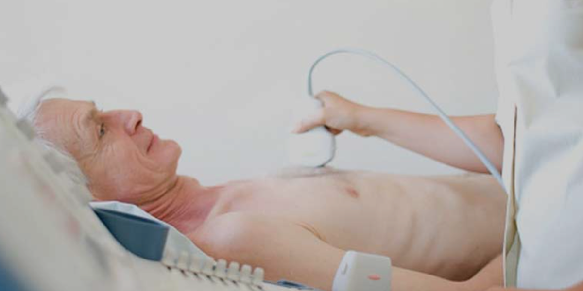
In the ever-evolving landscape of medical imaging, ultrasound stands out for its non-invasive nature, safety, and versatility. However, traditional two-dimensional ultrasound imaging, despite its widespread utility, offers limited perspectives. Enter ultrasound volume rendering techniques (VRT), an advanced development that transcends these limitations, offering a three-dimensional (3D) view that dramatically enhances diagnostic capabilities and treatment planning. This comprehensive exploration delves into the intricacies of ultrasound volume rendering, elucidating its principles, applications, benefits, and the future direction of this transformative technology.
The Essence of Ultrasound Volume Rendering Techniques
Volume rendering in ultrasound imaging is a sophisticated process that converts 2D echo data into 3D or 4D (3D plus time) visual representations. This technique relies on accumulating volumetric data sets from traditional ultrasound scans and applying complex algorithms to project these data into a 3D space, allowing for real-time visualization of internal structures.
How It Works
The journey from raw ultrasound data to a rendered volume begins with the acquisition of multiple cross-sectional images at various angles. These images are then compiled into a single, cohesive data set. Volume rendering software algorithms process this data, manipulating factors like opacity, brightness, and contrast to highlight different tissue types and structures within the volume. The result is a detailed, interactive 3D model that can be examined from any angle, providing unparalleled insight into the body’s internal architecture.
Clinical Applications and Benefits
Enhanced Diagnostic Accuracy
The most immediate benefit of ultrasound volume rendering is its ability to improve diagnostic accuracy. By offering a 3D representation, clinicians can better understand the spatial relationships and dimensions of anatomical structures and pathologies, significantly reducing interpretative errors.
Obstetrics and Gynecology
In obstetrics, VRT enables detailed examination of the fetal anatomy, aiding in the early detection of congenital anomalies. Similarly, in gynecology, it facilitates the evaluation of uterine and ovarian pathologies, improving the assessment of conditions like fibroids or polycystic ovarian syndrome.
Cardiology
Cardiac ultrasound with volume rendering provides a dynamic view of the heart and surrounding vessels. It assists in evaluating heart defects, valvular diseases, and other cardiovascular conditions with higher precision.
Surgery and Interventional Procedures
For surgeons, the ability to visualize and interact with a patient’s anatomy in three dimensions before and during procedures significantly enhances surgical planning and guidance. This is especially true for complex surgeries, where understanding the precise location and extent of pathology is critical.
Patient Communication
Ultrasound volume rendering also plays a vital role in patient education and engagement. By presenting intuitive and understandable images, patients can better grasp their conditions, fostering a collaborative approach to treatment planning.
Challenges and Future Directions
Despite its benefits, ultrasound volume rendering is not without challenges. The technique requires sophisticated hardware and software, as well as significant expertise in both acquiring and interpreting volumetric ultrasound data. Additionally, the quality of the rendered volume can be affected by patient movement and the inherent limitations of ultrasound, including dependency on operator skill and variations in tissue echogenicity.
However, the future of ultrasound volume rendering is bright, with ongoing advancements in computing power, algorithm efficiency, and integration with artificial intelligence poised to address these challenges. Researchers are continually developing ways to improve image quality, reduce processing times, and enhance the usability of this technology.
Conclusion
Ultrasound volume rendering techniques represent a quantum leap forward in the field of diagnostic imaging, offering a depth of detail and spatial awareness unmatched by conventional methods. As this technology continues to evolve, its integration into clinical practice is set to expand, promising to elevate the standards of patient care across a broad spectrum of medical disciplines. With each advancement, we move closer to unlocking the full potential of ultrasound imaging, illuminating the unseen complexities of the human body with ever-greater clarity and precision.