Summary
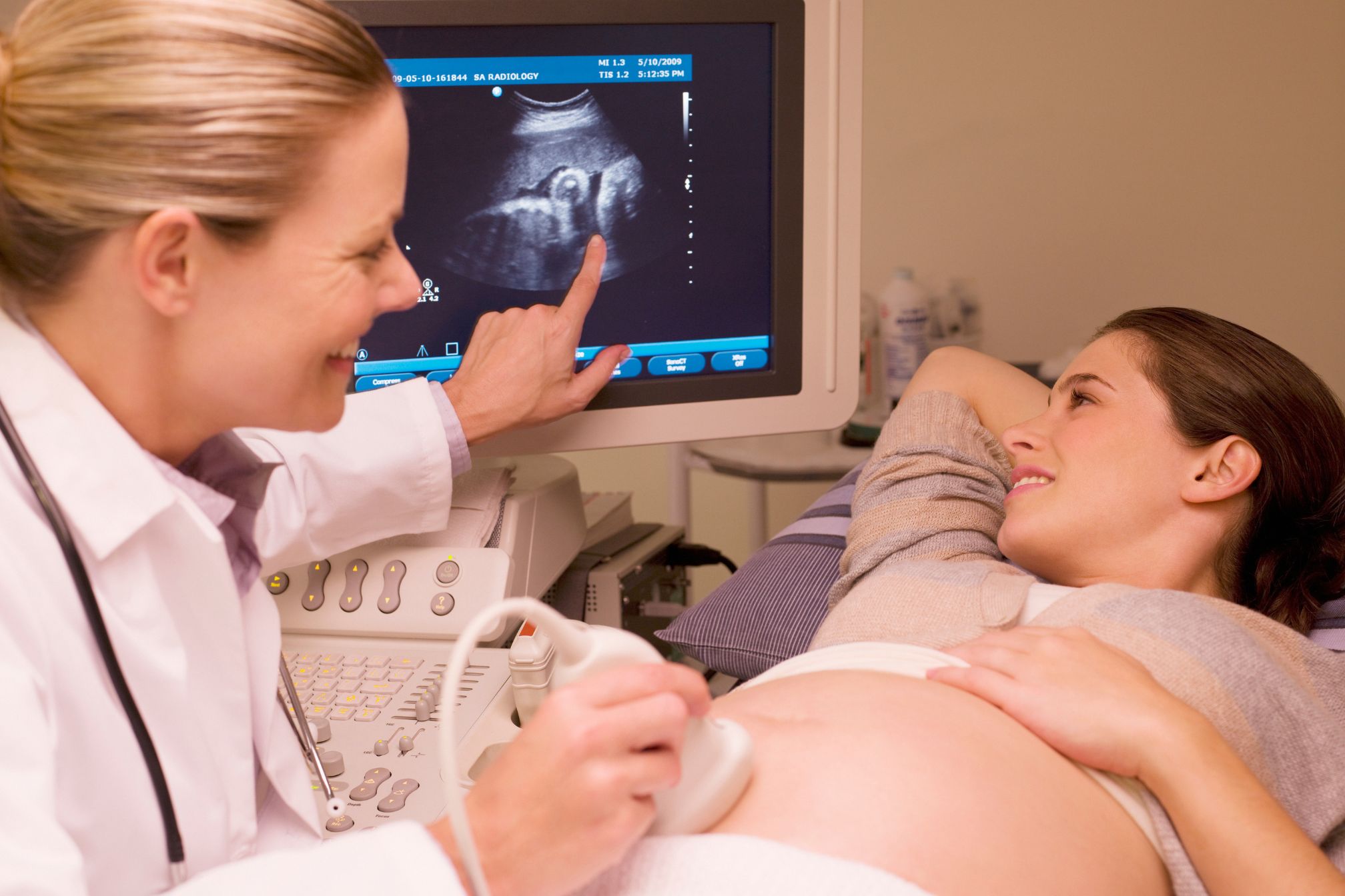
Ultrasound imaging is a powerful diagnostic tool used in many medical fields that provides real-time, non-invasive imaging of internal structures. Optimizing the ultrasound image is very important to ensure the quality of captured images, which helps in accurate diagnosis and patient management. In this article, we will explore the main methods of image optimization in relation to ultrasound.
Adjustment of Earnings and Time Gain Compensation (TGC):
Ultrasound systems allow sonographers to adjust the gain, which controls the brightness of the image. Correct gain settings are important for optimal contrast. In addition, the TGC settings allow compensation for the attenuation of sound waves at different depths, which ensures consistent image quality across the entire field of view.
Choosing the right sensor and frequency:
Different sensors offer different frequencies, and the choice of sensor and frequency depends on the type of study and the depth of the structures being imaged. High-frequency transducers provide better resolution of superficial structures, while low-frequency transducers penetrate deeper to image organs at greater depths.
Optimization for Color Doppler:
In cases where it is necessary to assess blood flow, such as vascular and cardiac ultrasound, optimizing color Doppler settings is crucial. Color gain, scale, and pulse repetition rate (PRF) adjustments are required to accurately visualize and measure blood flow velocity and direction.
Focus and depth settings:
Rapid focus of ultrasound is important to obtain clear images of specific areas of interest. Depth settings allow sonographers to control imaging depth and ensure that the region of interest is correctly imaged without unnecessary deep tissue disturbance.
Image post-processing:
Modern ultrasound equipment offers a variety of post-processing options that can improve image quality. These options include filtering, spatial fusion, and tissue harmonic imaging. Correct use of these features can improve image clarity.
Patient position and ultrasound technique:
Correct patient position is essential to obtain an optimal image. Sonographers must also ensure that they use appropriate techniques to minimize artifacts and improve image quality. This includes keeping a steady hand during imaging and considering the patient’s comfort.
Documentation and storage:
Effective storage and management of ultrasound images are essential. Picture archiving and communication systems (PACS) enable the organized storage, retrieval, and sharing of ultrasound images, ensuring their availability for comparison and diagnosis.
In conclusion, image optimization in ultrasound is a multifaceted process that requires a combination of technical expertise, device knowledge, and attention to detail. The application of these technologies ensures that the resulting images are diagnostically valuable, helping healthcare professionals provide accurate and efficient patient care.