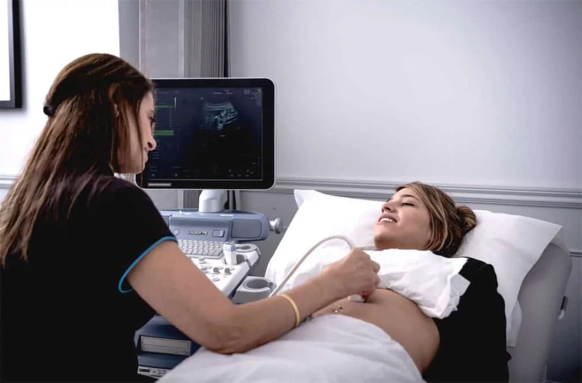
Ultrasound in Emergency Care: Rapid Diagnostic Techniques
Ultrasound is a crucial diagnostic tool in emergency care, providing immediate insights that assist in quick decision-making and treatment. Portable, non-invasive, and increasingly precise, ultrasound is now standard in emergency departments worldwide. Its rapid diagnostic capabilities allow healthcare providers to quickly assess internal injuries, determine the severity of illnesses, and guide lifesaving interventions.
This article explores the most effective ultrasound techniques for emergency diagnostics, highlighting how they contribute to faster, more accurate care.
Why Ultrasound is Essential in Emergency Care
In emergency settings, time is of the essence. Ultrasound’s portability and real-time imaging make it an ideal diagnostic modality when prompt decisions are critical. Unlike other imaging modalities like CT scans or MRIs, ultrasound machines can be used directly at the patient’s bedside, making them invaluable in urgent cases. Furthermore, ultrasound does not require ionizing radiation, making it safe for patients of all ages, including pregnant women and children.
Key Ultrasound Techniques in Emergency Diagnostics
- Focused Assessment with Sonography for Trauma (FAST)The FAST exam is one of the most frequently used ultrasound techniques in trauma cases. It is a rapid diagnostic tool used to detect free fluid, often a sign of internal bleeding, in critical areas such as the abdomen, pelvis, and chest. The FAST exam allows healthcare providers to assess areas where blood may pool, helping to identify injuries in patients who are hemodynamically unstable. It’s particularly useful for evaluating blunt abdominal trauma, helping clinicians to determine whether immediate surgical intervention is necessary.
- Extended FAST (eFAST)Building on the traditional FAST exam, the eFAST exam includes additional views to assess for pneumothorax (collapsed lung) and hemothorax (accumulation of blood in the pleural cavity). This expansion allows emergency practitioners to quickly evaluate life-threatening injuries to the thoracic cavity, which can be particularly beneficial in cases of chest trauma.
- Cardiac UltrasoundCardiac ultrasound, or echocardiography, is crucial in assessing patients with suspected cardiac issues, such as chest pain or heart failure. In emergency settings, a point-of-care echocardiogram can help determine the cause of chest pain, evaluate heart function, and detect conditions like pericardial effusion (fluid around the heart). A rapid cardiac assessment can provide information on the heart’s contractility, the presence of any valve abnormalities, and overall cardiac function, helping guide timely interventions.
- Lung UltrasoundLung ultrasound is increasingly utilized in emergency care due to its accuracy in diagnosing pulmonary conditions. It can be used to detect conditions like pneumothorax, pulmonary edema, pleural effusion, and pneumonia. In the context of respiratory distress, lung ultrasound can help determine the underlying cause, such as fluid overload or lung infection, enabling healthcare providers to start appropriate treatment immediately.
- Aorta UltrasoundA ruptured or dissected aorta is a life-threatening condition that requires immediate attention. Aorta ultrasound, often performed in patients presenting with severe abdominal or back pain, can detect aortic aneurysms and dissections. A quick, accurate assessment allows for timely intervention, as aortic ruptures require rapid transfer to surgical care.
- Gallbladder and Abdominal UltrasoundPatients with right upper quadrant pain or symptoms of jaundice may undergo a gallbladder ultrasound to check for gallstones or inflammation of the gallbladder. Abdominal ultrasound can also help diagnose appendicitis, intestinal obstruction, or other abdominal issues, which is vital in emergency settings where quick diagnosis can alleviate pain and prevent complications.
- Pelvic Ultrasound in Obstetrics and Gynecology EmergenciesFor women of childbearing age presenting with abdominal pain, pelvic ultrasound can identify gynecological issues such as ectopic pregnancy, ovarian cysts, or pelvic inflammatory disease. Early diagnosis of ectopic pregnancy is essential, as it can be life-threatening if untreated. Ultrasound is also useful in monitoring fetal health in pregnant patients, identifying any complications, and determining next steps.
- Vascular UltrasoundVascular ultrasound, such as Doppler ultrasound, is commonly used to evaluate blood flow in cases of suspected deep vein thrombosis (DVT). DVT can lead to pulmonary embolism, a potentially fatal condition. Using ultrasound to diagnose vascular issues allows for timely anticoagulant therapy and reduces the risk of further complications.
Benefits of Ultrasound in Emergency Medicine
The benefits of ultrasound in emergency care extend beyond rapid imaging and diagnostic capabilities. Here are some key advantages:
- Non-Invasive: Ultrasound is a non-invasive technique, reducing the risk of complications and minimizing patient discomfort.
- Real-Time Imaging: Real-time imaging allows practitioners to make immediate decisions based on current data, particularly important in life-threatening situations.
- Cost-Effective: Compared to other imaging methods, ultrasound is relatively cost-effective and can reduce the need for more expensive diagnostics like CT or MRI.
- Portable: The portability of ultrasound devices allows providers to use them directly at the patient’s bedside, making it ideal for emergency situations or pre-hospital settings.
Challenges and Limitations
Despite its advantages, ultrasound does have some limitations. Image quality can be affected by the patient’s body habitus or by the presence of excessive gas in the abdomen, which can obscure imaging. Additionally, accurate interpretation of ultrasound images requires skill and experience, which can limit its effectiveness if performed by less trained personnel. Continuous training and the availability of qualified ultrasound technicians are essential to maximize its utility in emergency care.
Best Practices for Using Ultrasound in Emergency Settings
To optimize ultrasound use in emergency care, healthcare providers should consider these best practices:
- Training and Skill Development: Regular training sessions and skill assessments can improve the diagnostic accuracy of ultrasound. Providers should stay updated on new techniques and guidelines in emergency ultrasound applications.
- Collaboration and Communication: Clear communication among team members in the emergency department can enhance the effectiveness of ultrasound. Providers should collaborate closely to ensure rapid interpretation and action based on ultrasound findings.
- Integration with Other Diagnostics: While ultrasound is powerful, it should be used alongside other diagnostics when possible. In some cases, CT scans or MRIs may be necessary to confirm initial ultrasound findings.
FAQ
Why is ultrasound important in emergency care?
Ultrasound provides quick, real-time imaging, allowing healthcare providers to make immediate decisions in critical situations.
What is a FAST exam, and when is it used?
The FAST exam is a rapid ultrasound test used in trauma cases to detect internal bleeding in areas like the abdomen and chest.
How does lung ultrasound benefit patients with respiratory distress?
Lung ultrasound can quickly diagnose conditions like pneumothorax, pulmonary edema, and pneumonia, aiding in targeted treatment.
What does an eFAST exam include that a FAST exam doesn’t?
The eFAST exam includes additional views to assess for pneumothorax and hemothorax, helping detect chest injuries.
How can ultrasound detect an aortic aneurysm?
Aorta ultrasound can visualize the aorta, helping identify aneurysms or dissections that require immediate intervention.
Why is cardiac ultrasound useful in emergency settings?
Cardiac ultrasound assesses heart function and can detect conditions like pericardial effusion, providing rapid insight into cardiac health.
What is the role of ultrasound in gynecological emergencies?
Pelvic ultrasound helps diagnose ectopic pregnancy, ovarian cysts, and other gynecological conditions in women with abdominal pain.
How is Doppler ultrasound used in vascular emergencies?
Doppler ultrasound assesses blood flow, helping diagnose deep vein thrombosis and reduce the risk of pulmonary embolism.
What are some benefits of ultrasound over other imaging methods?
Ultrasound is non-invasive, portable, cost-effective, and provides real-time imaging without ionizing radiation.
What are the limitations of ultrasound in emergency care?
Limitations include poor imaging in certain body types and the need for trained personnel for accurate interpretation.
Conclusion
Ultrasound is an invaluable tool in emergency medicine, offering rapid, non-invasive diagnostics that save time and lives. Techniques like the FAST exam, cardiac and lung ultrasounds, and aorta imaging allow healthcare providers to diagnose critical conditions quickly and accurately. By providing real-time insights at the point of care, ultrasound empowers emergency physicians to make prompt, informed decisions that significantly improve patient outcomes. As ultrasound technology continues to evolve, its role in emergency care will likely expand further, offering new diagnostic capabilities and enhancing the quality of urgent medical interventions.


