The evolution of medical imaging has reached an exciting juncture with the advent of three-dimensional (3D) carotid ultrasound imaging, marking a significant leap in diagnosing and managing vascular conditions. This groundbreaking technology has transformed our approach to visualizing the intricate structures of the carotid arteries, offering unparalleled detail and accuracy. By providing a comprehensive three-dimensional view, 3D carotid ultrasound imaging enriches our understanding of vascular pathologies and enhances patient care. This article delves into the advancements, applications, benefits, and future prospects of 3D carotid ultrasound imaging in clinical practice.
The Evolution of Carotid Imaging
The carotid arteries, vital conduits supplying blood to the brain, are common focal points for atherosclerotic disease, which can lead to life-threatening conditions such as strokes. Traditional two-dimensional (2D) ultrasound has been instrumental in evaluating carotid artery disease, providing valuable information about blood flow, plaque presence, and stenosis. However, the introduction of three-dimensional ultrasound technology has revolutionized this field, offering a more nuanced and comprehensive view of these crucial vessels.
Principles of 3D Carotid Ultrasound Imaging
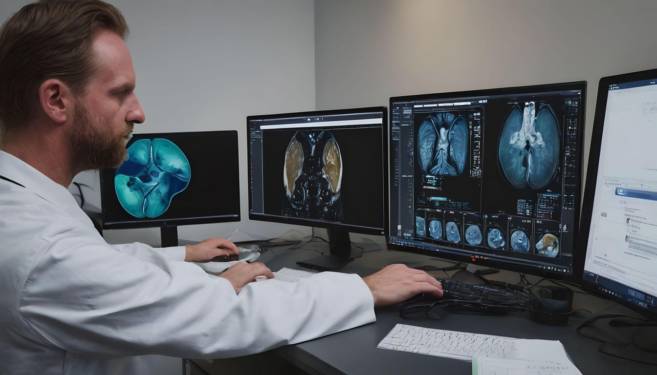
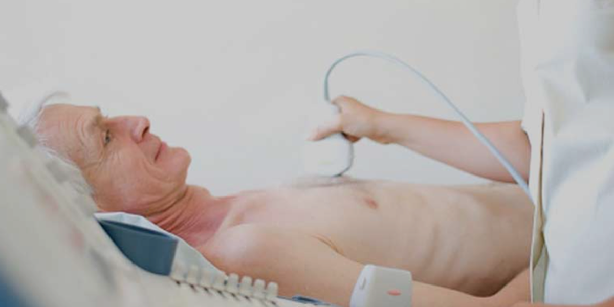
3D carotid ultrasound imaging utilizes sophisticated transducers to emit sound waves that capture detailed cross-sectional images of the carotid arteries from multiple angles. Advanced software algorithms then compile these images into a cohesive 3D model, allowing clinicians to observe the artery’s structure in its entirety. This method not only illuminates the vessel’s lumen but also provides insight into the arterial wall and any atherosclerotic plaque that may be present.
Key Applications and Advantages
Enhanced Visualization of Plaque Composition
One of the most significant advantages of 3D carotid ultrasound imaging is its ability to visualize plaque composition with high precision. The technology distinguishes between stable plaques, which are less likely to cause acute events, and vulnerable plaques, which pose a higher risk of rupture and subsequent stroke. This differentiation is critical in risk stratification and determining the appropriate management strategy for each patient.
Improved Assessment of Stenosis
3D imaging offers an improved assessment of the degree of stenosis, providing exact measurements of the arterial lumen. This accuracy is paramount in surgical planning for patients who may require carotid endarterectomy or stenting.
Superior Evaluation of Treatment Response
Following interventions, 3D carotid ultrasound imaging serves as an excellent tool for monitoring treatment efficacy, visualizing changes in plaque volume and composition over time, and guiding further therapeutic decisions.
The Future of 3D Carotid Imaging
The future of 3D carotid ultrasound imaging is promising, with ongoing advancements aimed at enhancing image resolution, reducing scanning time, and improving user-friendliness. Integration with artificial intelligence (AI) stands as a potential frontier, where AI algorithms could automate aspects of the imaging process, provide instant analyses of plaque characteristics, and predict the risk of future vascular events with higher accuracy. Additionally, the development of portable 3D ultrasound devices would further increase the accessibility of this technology, making comprehensive vascular assessments available in a wider range of clinical settings.
Challenges and Considerations
Despite its benefits, the widespread adoption of 3D carotid ultrasound imaging faces challenges. High costs, the need for specialized training to interpret 3D images accurately, and variability in image quality due to patient factors are among the primary concerns. Moreover, establishing standardized protocols for 3D imaging and analysis is crucial to ensure consistency and reliability across different institutions.
Conclusion
Three-dimensional carotid ultrasound imaging embodies a significant advancement in vascular diagnostics, providing a depth of detail unmatched by traditional methods. As the technology continues to evolve, it holds the promise of not only enriching our understanding of vascular diseases but also pioneering personalized approaches to patient care. Despite existing challenges, the potential benefits of 3D carotid imaging in preventing strokes and improving patient outcomes are undeniable. Navigating the new dimensions offered by this technology will undoubtedly play a pivotal role in the future of cardiovascular medicine.