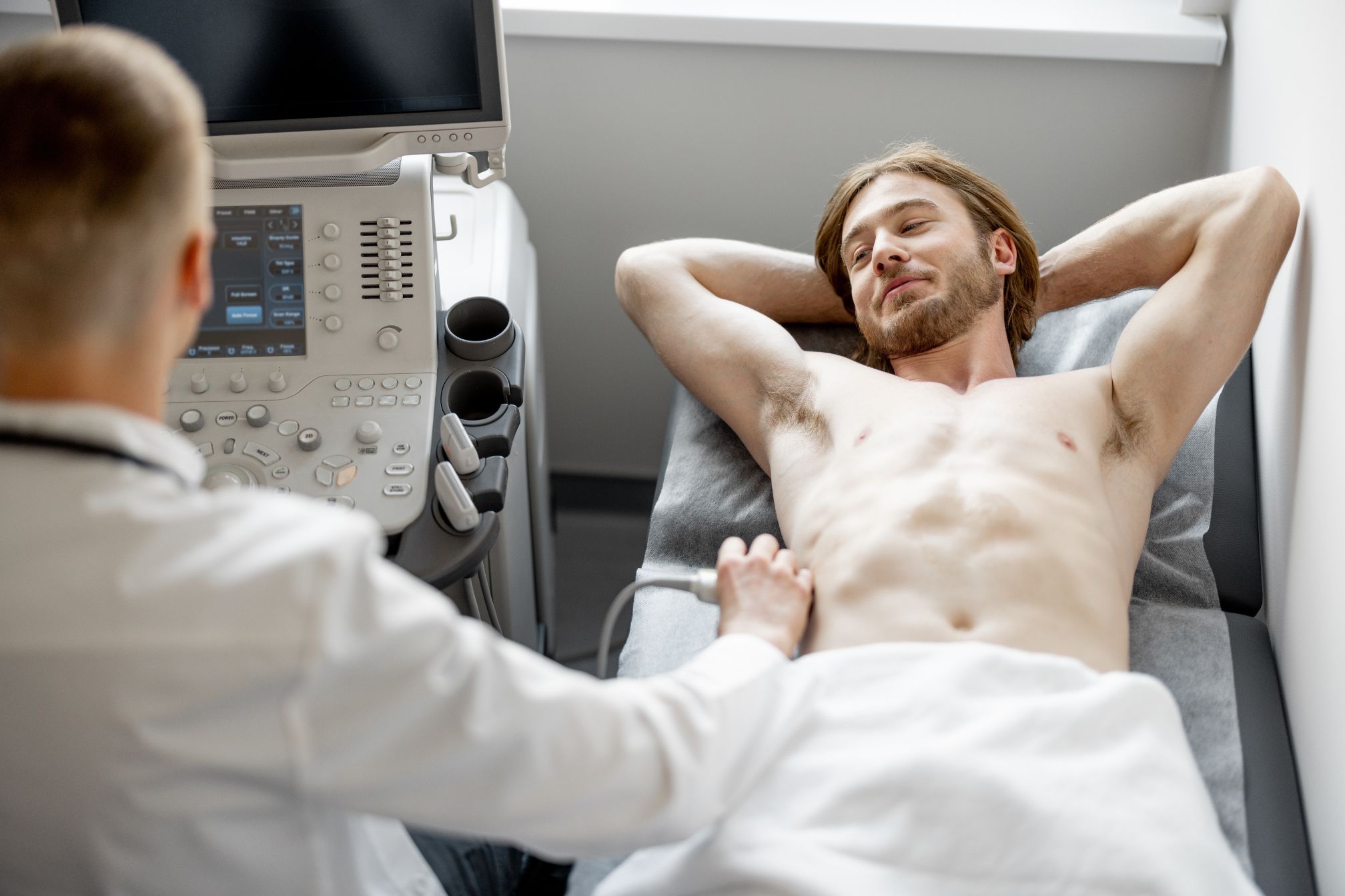
When it comes to Ultrasound Diagnosis of Lymph Nodes the power of sound waves is nothing short of revolutionary. Picture this: You’re at the doctor’s office, feeling a bit anxious about that lump you found under your arm. Instead of diving straight into invasive tests, your doctor suggests an ultrasound. In just a few minutes, with a little gel and a handheld device, you’re able to get a detailed look inside your body—all without a single incision. That’s the magic of ultrasound.
Why Ultrasound Diagnosis of Lymph Nodes?
So, why Ultrasound Diagnosis of Lymph Nodes becoming the go-to method for doctors? It’s simple. Ultrasound is non-invasive, meaning there’s no need to break the skin or use any needles—unless you need a biopsy, which ultrasound can also guide with pinpoint accuracy. Remember the last time you had an X-ray or MRI? Those machines can be a bit intimidating, not to mention the radiation exposure with X-rays. Ultrasound sidesteps all that. It’s completely safe and painless. The process is as easy as lying down, applying a bit of cool gel, and letting the transducer do its thing.
The Science Behind Ultrasound Diagnosis of Lymph Nodes
The beauty of Ultrasound Diagnosis of Lymph Nodes lies in its ability to use sound waves to create images. These sound waves bounce off the lymph nodes and surrounding tissues, creating detailed images that can tell us a lot about what’s happening inside.
Real-Time Results
One of the standout features of ultrasound is the ability to get real-time images. Imagine watching a live video of your lymph nodes as the doctor moves the transducer over your skin. This live feedback allows for immediate analysis and decision-making, which is crucial when time is of the essence.
From Suspicion to Clarity: Real-Life Example
Let’s dive into a real-life scenario. Jane, a 45-year-old mother of two, noticed a lump in her neck. Her first thought? Cancer. But instead of jumping to conclusions, her doctor suggested an ultrasound. Within minutes, they could see that the lymph node was just reactive—a harmless response to a recent cold. No biopsy needed, no unnecessary panic. This is the kind of clarity that Ultrasound Diagnosis of Lymph Nodes can bring.
The Versatility of Ultrasound in Lymph Node Assessment
When we talk about Ultrasound Diagnosis of Lymph Nodes we’re not just talking about cancer detection. Ultrasound is incredibly versatile and can be used to assess lymph nodes for a variety of reasons.
Cancer Staging and Monitoring
For patients already diagnosed with cancer, ultrasound is an invaluable tool. It helps doctors stage the cancer by determining whether it has spread to the lymph nodes. Moreover, it’s used to monitor lymph nodes during and after treatment, ensuring that the cancer hasn’t returned or spread.
Infections and Inflammatory Conditions
Lymph nodes can swell due to infections or inflammatory conditions like sarcoidosis. Ultrasound helps distinguish these from more sinister causes, guiding treatment decisions. For instance, a lymph node with a benign appearance on ultrasound might just need a course of antibiotics, while one with suspicious features might warrant further investigation.
What Makes Ultrasound So Accurate?
Accuracy in Ultrasound Diagnosis of Lymph Nodes isn’t just about the images themselves. It’s about how those images are interpreted and used.
Detailed Images
Modern ultrasound machines are equipped with high-resolution capabilities, providing detailed images that allow doctors to see the size, shape, and internal structure of lymph nodes. This detail is crucial for distinguishing between benign and malignant conditions.
Assessment of Vascularity
Another critical aspect is the assessment of vascularity using Doppler ultrasound. By evaluating blood flow within the lymph nodes, doctors can identify patterns that may indicate malignancy. For example, increased blood flow is often associated with cancerous nodes, helping in early detection and intervention.
Guiding Precision with Ultrasound-Guided Biopsy
When a lymph node looks suspicious, the next step might be a biopsy. Here’s where Ultrasound Diagnosis of Lymph Nodes shines even brighter. Ultrasound can guide the biopsy needle with such precision that it minimizes discomfort and maximizes accuracy.
Fine-Needle Aspiration (FNA) and Core Needle Biopsy (CNB)
These are the most common types of biopsies done with ultrasound guidance. FNA uses a thin needle to extract cells, while CNB uses a larger needle to remove a small tissue sample. Both methods are minimally invasive and highly accurate, thanks to the real-time guidance of ultrasound.
The Future of Ultrasound in Lymph Node Diagnosis
As technology continues to evolve, the role of Ultrasound Diagnosis of Lymph Nodes is set to expand. We’re talking about even more precise imaging, better differentiation between benign and malignant nodes, and the potential for artificial intelligence to assist in interpretation.
Artificial Intelligence Integration
Imagine a future where AI analyzes your ultrasound in real-time, providing immediate feedback to your doctor. This could enhance accuracy and speed up the diagnostic process, getting patients the treatment they need faster.
FAQ
What will an ultrasound of lymph nodes show?
An ultrasound of lymph nodes reveals their size, shape, internal structure, and vascularity. This helps differentiate between normal, reactive, and potentially malignant nodes, guiding further diagnosis and treatment.
What is the diagnostic test for lymph nodes?
The primary diagnostic test for lymph nodes includes ultrasound, often followed by a biopsy if abnormalities are detected. Other tests may include CT scans, MRI, or PET scans depending on the clinical scenario.
What are the ultrasound features of suspicious lymph nodes?
Suspicious lymph nodes on ultrasound may appear with irregular shapes, loss of the fatty hilum, increased blood flow, and a more heterogeneous or hypoechoic texture. These features often prompt further investigation.
Can an ultrasound detect lymph cancer?
Yes, an ultrasound can detect signs of lymph cancer by identifying suspicious characteristics in the lymph nodes, such as abnormal size, shape, and vascularity. However, a biopsy is usually required for a definitive diagnosis.
How accurate is a lymph node ultrasound?
Lymph node ultrasounds are highly accurate, especially when combined with Doppler imaging and guided biopsies. They are effective in distinguishing between benign and malignant nodes, though the accuracy can vary based on the operator’s skill.
How to tell if a lymph node is cancerous?
A cancerous lymph node may present as enlarged, irregularly shaped, with a loss of the central fatty hilum, and increased blood flow on ultrasound. A biopsy is required for confirmation.
What is the diagnosis for enlarged lymph node?
The diagnosis for an enlarged lymph node can vary, ranging from benign reactive changes due to infection to more serious conditions like cancer. Ultrasound and biopsy are key tools in determining the cause.
What are the characteristics of a malignant lymph node?
Malignant lymph nodes are often irregular in shape, larger than normal, with a loss of the central fatty hilum, and may show increased internal blood flow on Doppler ultrasound.
Are lymph nodes painful?
Lymph nodes can be painful, especially if they are swollen due to infection or inflammation. However, malignant lymph nodes are often painless, which is why further evaluation is important if a node feels abnormal.
Conclusion
In the world of medical diagnostics, Ultrasound Diagnosis of Lymph Nodes stands out as a game-changer. Its ability to provide quick, accurate, and non-invasive assessments makes it an indispensable tool for doctors and a reassuring option for patients. Whether you’re worried about a lump or monitoring a known condition, ultrasound offers a clear, safe, and effective path to answers. And as the technology continues to improve, its role in healthcare will only grow, bringing even more benefits to patients around the world