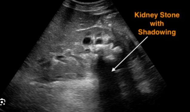
This ultrasound image shows a kidney stone with acoustic shadowing. Let’s break down what we see:
- Kidney Stone: The bright hyperechoic (white) spot indicated by the arrow is the kidney stone. Its high density causes it to reflect the ultrasound waves strongly, making it appear bright on the image.
- Acoustic Shadowing: The dark area behind the stone is the acoustic shadow. This occurs because the stone blocks the ultrasound waves, preventing them from reaching the tissues behind it. This shadowing is a key feature in identifying kidney stones on ultrasound.
- Surrounding Tissue: The surrounding tissue appears in varying shades of gray, representing different densities and structures within the kidney.
- The size and location of the stone can be determined from the ultrasound image.
- Further tests, such as a CT scan, may be needed to confirm the diagnosis and plan treatment.
- Treatment for kidney stones varies depending on the size and location of the stone and may include medication, shock wave lithotripsy, or surgery.
No related posts.