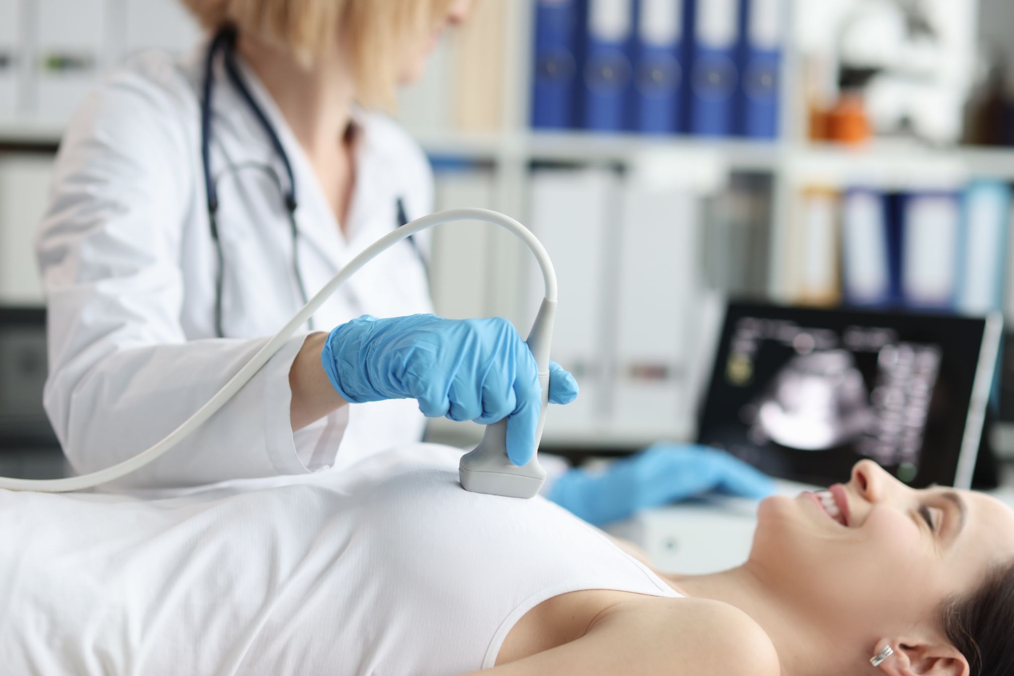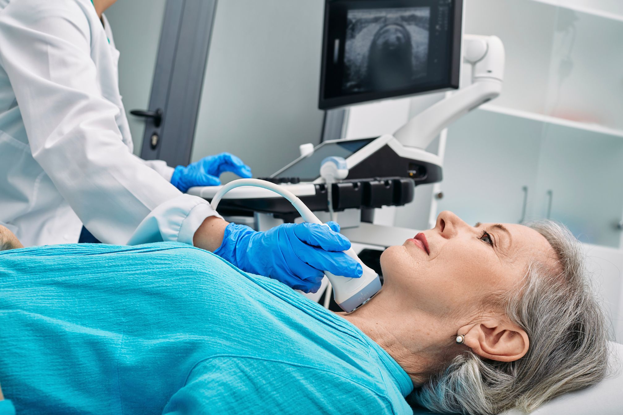
When it comes to diagnosing thyroid conditions, the ultrasound in thyroid mucosa-associated lymphoid tissue plays a pivotal role. This imaging technique provides invaluable insights into the health of the thyroid gland, especially when dealing with conditions associated with mucosa-associated lymphoid tissue (MALT). Let’s dive into how ultrasound can illuminate this complex area and why it’s such an essential tool in modern medicine.
What Is Thyroid Mucosa-Associated Lymphoid Tissue?
Before we get into the specifics of ultrasound, it’s crucial to understand what MALT is and its role in the thyroid. Mucosa-associated lymphoid tissue is a type of lymphoid tissue found in mucosal surfaces throughout the body, including the thyroid. In the thyroid, MALT can be involved in several conditions, from autoimmune diseases to lymphomas. MALT in the thyroid is critical because it can indicate various thyroid disorders. Conditions like autoimmune thyroiditis and thyroid lymphomas often involve changes in this tissue. Detecting and monitoring these changes is where ultrasound becomes incredibly valuable.
How Ultrasound Helps in Assessing Thyroid MALT
1. Detecting Thyroid Nodules
One of the primary uses of ultrasound in thyroid MALT evaluation is the detection of nodules. Thyroid nodules can be benign or malignant, and distinguishing between them is essential. Ultrasound allows for the identification of these nodules, often highlighting their size, shape, and internal characteristics.
For example, imagine a patient who has been feeling a lump in their throat. An ultrasound can quickly identify whether this lump is a thyroid nodule associated with MALT or something else entirely. By providing clear images, ultrasound helps doctors make accurate diagnoses.
2. Characterizing Lesions
Once a nodule is detected, the next step is to characterize it. Ultrasound can determine whether a nodule is solid or cystic, its echogenicity, and its margins. These features are crucial for assessing the potential malignancy of the nodule.
For instance, a solid nodule with irregular borders and increased vascularity might raise red flags for malignancy. On the other hand, a smooth, well-defined nodule might be benign. Ultrasound provides these insights in real time, allowing for prompt decision-making.
3. Monitoring Disease Progression
Ultrasound isn’t just for diagnosis; it’s also used to monitor disease progression. For patients with thyroid MALT disorders, regular ultrasound exams can track changes in nodules or lesions over time.
Consider a patient undergoing treatment for autoimmune thyroiditis. Ultrasound can show how the disease is responding to therapy, such as whether nodules are shrinking or becoming more stable. This ongoing monitoring helps adjust treatment plans and ensures the best possible outcome.
Real-Life Applications of Ultrasound in Thyroid MALT Disorders
1. Thyroid Lymphoma: An Inside Look
Thyroid lymphoma is a type of cancer that affects the thyroid gland and is often associated with MALT. Ultrasound plays a crucial role in diagnosing and monitoring this condition. Typically, a thyroid lymphoma appears as a hypoechoic mass with irregular margins on ultrasound. Increased vascularity might also be observed.
Take, for example, a patient with a suspected thyroid lymphoma. The ultrasound might reveal a mass with features consistent with lymphoma, guiding the next steps for biopsy and treatment. This early detection can significantly impact the effectiveness of the treatment plan.
2. Autoimmune Thyroiditis: Spotting the Signs
Autoimmune thyroiditis, including Hashimoto’s thyroiditis, involves inflammation of the thyroid gland. Ultrasound can reveal diffuse hypoechogenicity and increased vascularity in the thyroid tissue, helping to diagnose and assess the extent of the disease.
Imagine a scenario where a patient presents with symptoms of thyroiditis. An ultrasound can confirm the diagnosis by showing characteristic changes in the thyroid gland, such as a heterogeneous echotexture. This helps in understanding the disease’s progression and tailoring appropriate treatment.
3. MALT Lymphoma: Detailed Imaging
MALT lymphoma can present in various ways on ultrasound, from focal lesions to diffuse changes. The appearance can range from hypoechoic to heterogeneous, depending on the disease stage and characteristics.
Consider a case where a patient’s ultrasound shows a “moth-eaten” appearance of the thyroid tissue. This could indicate MALT lymphoma, prompting further investigation and treatment. The detailed imaging provided by ultrasound is crucial for accurately diagnosing and managing this condition.
Limitations and Considerations of Ultrasound
While ultrasound is a powerful tool, it’s not without limitations. For instance, it might not always differentiate between MALT-related thyroid conditions and other thyroid disorders. In some cases, additional imaging techniques like CT or MRI may be needed for a comprehensive evaluation.
Moreover, the resolution of ultrasound can vary, which might affect the detection of small or subtle changes in thyroid MALT. Despite these limitations, ultrasound remains a fundamental tool in the assessment and management of thyroid MALT disorders.
FAQ
What is a thyroid ultrasound for lymph nodes?
A thyroid ultrasound for lymph nodes uses sound waves to create images of the thyroid gland and surrounding lymph nodes. This imaging technique helps in evaluating abnormalities such as swelling or suspicious masses in the lymph nodes.
Can thyroid lymphoma be seen on ultrasound?
Yes, thyroid lymphoma can be detected on ultrasound. It typically appears as a hypoechoic mass with irregular borders and increased blood flow, helping doctors assess the presence and extent of the lymphoma.
What is mucosa associated with lymphoid tissue?
Mucosa-associated lymphoid tissue (MALT) is a type of lymphoid tissue found in mucosal surfaces throughout the body. In the thyroid, MALT can play a role in various disorders, including autoimmune conditions and lymphomas.
Is mucosa associated lymphoid tissue cancer?
Mucosa-associated lymphoid tissue (MALT) itself is not cancerous, but it can be involved in MALT lymphoma, a type of cancer. This lymphoma affects the lymphoid tissue in mucosal areas, including the thyroid.
How does an ultrasound tell if a lymph node is cancerous?
Ultrasound evaluates lymph nodes for cancer by assessing their size, shape, and internal characteristics. Irregular margins, increased vascularity, and changes in texture may suggest malignancy and warrant further investigation.
What is the purpose of a thyroid ultrasound?
The purpose of a thyroid ultrasound is to provide detailed images of the thyroid gland. It helps in diagnosing conditions like nodules, cysts, and tumors, and in monitoring the progression of thyroid diseases and assessing treatment responses.
What happens after a thyroid ultrasound?
After a thyroid ultrasound, the images are analyzed by a radiologist who provides a report to your doctor. Your doctor will review the findings to determine if any further tests or treatments are needed based on the ultrasound results.
What are red flags on thyroid ultrasound?
Red flags on a thyroid ultrasound include irregularly shaped nodules, increased vascularity, microcalcifications, and uneven echogenicity. These features may indicate a higher risk of malignancy and require additional evaluation.
What does a normal thyroid ultrasound look like?
A normal thyroid ultrasound shows a smooth, homogeneous thyroid gland with no abnormal nodules or cysts. The tissue should appear uniformly echogenic, and the overall structure should be well-defined without irregularities or significant vascular changes
Conclusion
In summary, the role of ultrasound in thyroid mucosa-associated lymphoid tissue is both profound and multifaceted. From detecting and characterizing nodules to monitoring disease progression, ultrasound provides essential insights that guide diagnosis and treatment. Its real-time imaging capabilities and non-invasive nature make it an invaluable tool in the evaluation of thyroid conditions associated with MALT.
Whether it’s identifying a suspicious nodule, assessing the impact of autoimmune thyroiditis, or monitoring thyroid lymphoma, ultrasound continues to be a cornerstone of thyroid imaging. As technology advances, its applications and accuracy are likely to improve, further enhancing its role in managing thyroid MALT disorders