The liver is a key organ for metabolism, detoxification, and blood filtration, making its health a critical focus in medical care. Ultrasound liver scanning is a non-invasive and reliable diagnostic tool that is essential for assessing, diagnosing, and monitoring liver diseases. This article offers a comprehensive guide to ultrasound liver scanning, detailing its techniques, applications, and its pivotal role in effective healthcare management.
Introduction to Ultrasound Liver Scanning
Ultrasound liver scanning utilizes high-frequency sound waves to produce images of the liver. This method allows healthcare providers to visualize the liver’s structure, assessing its size, shape, and the presence of any lesions or abnormalities. Being non-invasive, ultrasound scanning is often preferred over other diagnostic techniques for initial liver assessment.
Preparing for a Liver Ultrasound Scan
Preparation for a liver ultrasound is minimal yet crucial for obtaining clear images. Patients are typically advised to fast for several hours (usually 6-8 hours) before the scan to reduce gas in the stomach and intestines, which can obstruct the view of the liver. Unlike more invasive procedures, there are no risks associated with fasting, and patients can return to their normal activities immediately after the scan.
The Ultrasound Liver Scanning Process
The liver ultrasound scanning process is straightforward and painless:
- Positioning: The patient is positioned on an examination table, usually lying on their back or slightly turned to the left side to expose the right upper quadrant of the abdomen, where the liver is located.
- Gel Application: A water-based gel is applied to the skin over the liver. This gel helps to conduct the ultrasound waves and eliminate air between the transducer (probe) and skin.
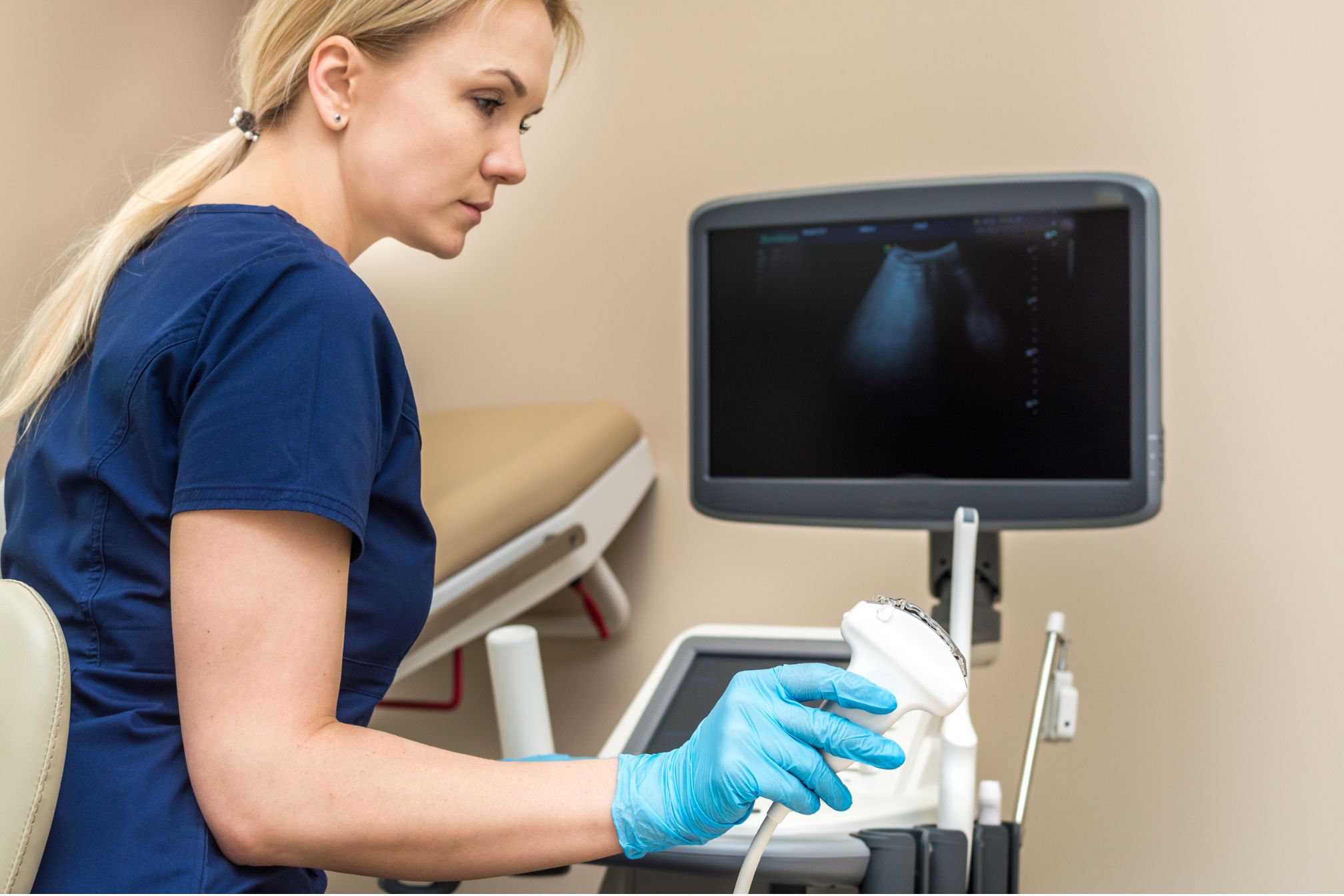
- Scanning: The sonographer (ultrasound technician) places the transducer against the skin and moves it around to capture images of the liver from various angles. The transducer emits sound waves that penetrate the body, bounce off the liver and other organs, and return to the transducer. The ultrasound machine analyzes these echoes to create images.
- Interpretation: The generated images allow the healthcare provider to evaluate the liver’s structure. The sonographer may take measurements and make notes during the scan, which are then reviewed by a radiologist or physician for diagnosis.
Applications of Liver Ultrasound Scanning
Liver ultrasound scanning has several critical applications in medical diagnostics and patient care:
- Detection of Liver Diseases: It helps in diagnosing a range of liver conditions, including fatty liver disease, cirrhosis, hepatitis, and liver cancer.
- Identification of Abnormalities: It can detect abnormalities such as cysts, tumors, and hemangiomas within the liver.
- Guided Procedures: Ultrasound serves as a guide for procedures like biopsies, helping to ensure accuracy and safety.
- Monitoring: It allows for the monitoring of known liver conditions, assessing disease progression or the effectiveness of treatments.
Advantages of Ultrasound Liver Scanning
- Non-Invasive: Unlike CT scans or MRIs, ultrasound doesn’t use ionizing radiation, making it safer for repeated use.
- Accessibility and Convenience: Ultrasound machines are widely available and portable, making liver scans accessible in various healthcare settings.
- Real-Time Imaging: It provides immediate results, which is crucial for diagnosing acute conditions or guiding interventional procedures.
Challenges and Limitations
While ultrasound liver scanning is highly effective, certain factors can limit its accuracy:
- Body Composition: In overweight or obese patients, the penetration of ultrasound waves can be hindered, affecting image quality.
- Interpretation Variances: The quality of the ultrasound images and the interpretation can vary depending on the skill of the sonographer and the quality of the ultrasound equipment.
Conclusion
Ultrasound liver scanning is a cornerstone in diagnosing and monitoring liver diseases, offering a blend of safety, accessibility, and diagnostic capability. As advancements in ultrasound technology continue, so too will its applications and effectiveness in liver disease management. This scanning technique not only facilitates early detection and intervention but also significantly contributes to patient care by guiding treatment strategies and monitoring disease progress. In the dynamic landscape of medical diagnostics, ultrasound liver scanning remains an invaluable asset, echoing the evolving nature of healthcare innovation.


