Sonographic imaging, commonly known as ultrasound imaging, is a cornerstone of modern medical diagnostics, offering real-time, non-invasive visualization of internal structures and organs. This imaging modality utilizes high-frequency sound waves to create images of tissues and organs inside the body, aiding in the diagnosis and management of various medical conditions. Understanding the essentials of sonographic imaging is crucial for both healthcare professionals and patients to appreciate its capabilities and applications.
How Sonographic Imaging Works
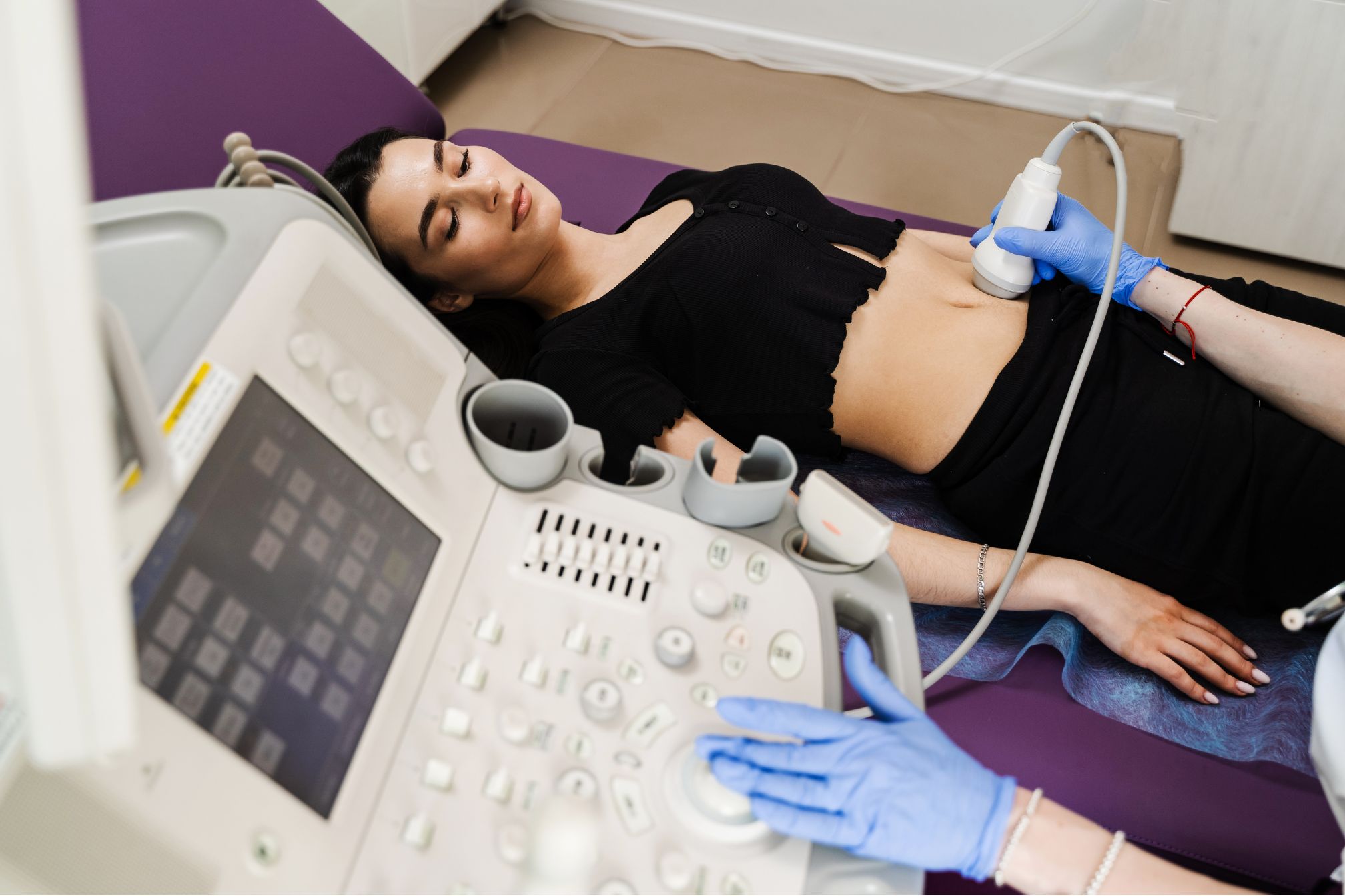
Sonographic imaging works on the principle of sound wave propagation and reflection. A transducer, which emits and receives sound waves, is placed on the skin surface and moved over the area of interest. These sound waves travel through the body and bounce off tissues with different acoustic properties. The echoes produced are then converted into electrical signals and processed by a computer to generate real-time images on a monitor.
Applications of Sonographic Imaging
Sonographic imaging is used across numerous medical specialties for diagnostic purposes:
- Obstetrics and Gynecology: It plays a vital role in monitoring fetal development during pregnancy, assessing the female reproductive system, and diagnosing conditions such as ovarian cysts or uterine fibroids.
- Cardiology: In echocardiography, ultrasound imaging helps evaluate the structure and function of the heart, detecting abnormalities like valve disorders or congenital heart defects.
- Abdominal Imaging: Ultrasound is used to examine the liver, gallbladder, pancreas, kidneys, and other abdominal organs. It helps diagnose conditions such as gallstones, liver cirrhosis, or kidney tumors.
- Musculoskeletal Imaging: Ultrasound aids in assessing soft tissues, muscles, tendons, and joints for injuries, inflammation, or abnormalities like tendon tears or arthritis.
- Vascular Imaging: Doppler ultrasound is employed to evaluate blood flow and detect conditions such as deep vein thrombosis (DVT), carotid artery disease, or peripheral arterial disease (PAD).
Advantages of Sonographic Imaging
Sonographic imaging offers several advantages over other imaging modalities:
- Non-invasive: It does not involve ionizing radiation, making it safer than X-rays or CT scans, especially for pregnant women and children.
- Real-time Imaging: Provides immediate visualization of moving structures and functions, such as heart valves or blood flow, aiding in dynamic assessments.
- Portable and Versatile: Ultrasound machines are portable, allowing for bedside or point-of-care use in hospitals, clinics, and even remote locations.
- Cost-effective: Generally less expensive than other imaging techniques, making it accessible for routine screenings and monitoring.
Challenges and Limitations
While highly valuable, sonographic imaging has its limitations:
- Operator Dependence: Image quality and diagnostic accuracy heavily rely on the operator’s skill and experience in transducer positioning and image interpretation.
- Obesity and Gas Interference: Body habitus, excessive adipose tissue, or bowel gas can hinder sound wave penetration, affecting image quality.
- Limited Depth and Resolution: Higher frequencies offer better resolution but have limited penetration depth, whereas lower frequencies penetrate deeper but provide less detailed images.
Future Directions
The future of sonographic imaging is promising with ongoing advancements:
- Artificial Intelligence: Integration of AI for automated image analysis, enhancing diagnostic accuracy and efficiency.
- Enhanced Imaging Techniques: Continued development of techniques like contrast-enhanced ultrasound, elastography, and 3D/4D imaging for improved tissue characterization and visualization.
- Miniaturization and Wireless Technology: Advancements in miniaturization and wireless connectivity will further enhance the portability and usability of ultrasound devices.
Conclusion
Sonographic imaging has revolutionized medical diagnostics by offering safe, real-time visualization of internal structures without the use of radiation. Understanding its essentials—from how it works to its applications across various medical fields—is crucial for healthcare professionals to leverage its capabilities effectively. As technology evolves, sonographic imaging continues to play a pivotal role in improving patient care, enhancing diagnostic accuracy, and expanding the scope of medical imaging capabilities.